Skeletal system · Head and neck
Sphenoid bone !
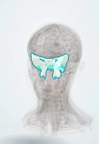
The sphenoid is a butterfly-shaped bone in the middle of the skull base; it links the skull bones and holds the pituitary gland in its central saddle.
Opens in the Durgical app, no sign-up needed.
What it is
A butterfly-shaped bone in the middle of the skull base; its central saddle holds the pituitary gland.
What it does
Links the skull bones together and lets many nerves and vessels pass through its openings.
Key facts
- Joints with
- Frontal, parietals, temporals, occipital, ethmoid, vomer, palatines, zygomatics
- Landmarks
- Sella turcica, greater and lesser wings, pterygoid processes, optic canal, superior orbital fissure, foramina rotundum, ovale and spinosum, sphenoid sinus; pterygoid (Vidian) canal; pterygoid hamulus (pulley of tensor veli palatini); spine of the sphenoid (sphenomandibular ligament)
Relations
- Above
- Pituitary gland in the sella turcica, with the optic chiasm above it; frontal lobes on the lesser wings and temporal lobes on the greater wings in the middle cranial fossa
- Lateral
- Cavernous sinuses against the body; temporalis and the infratemporal fossa against the outer surface of the greater wings
- In front
- Ethmoid bone and the back of the nasal cavity; the orbits, whose apex and posterior walls it forms
- Behind
- Basilar part of the occipital bone (clivus), joined at the spheno-occipital synchondrosis; the petrous parts of the temporal bones
- Below
- Roof of the nasopharynx; the pterygoid processes give origin to the medial and lateral pterygoid muscles, with the pterygopalatine fossa in front of them
How to find it
Only the greater wing comes near the surface, in the temple just behind the frontal process of the zygomatic bone, under temporalis. The rest is studied on CT and MRI and reached endoscopically through the nose and the sphenoid sinus.
On imaging
MRI of the sella (thin coronal and sagittal slices with contrast) shows the pituitary, the optic chiasm and the cavernous sinuses; CT with bone windows shows the foramina, the sinus septa and skull base fractures.
Clinical notes
- A pituitary macroadenoma growing up out of the sella presses on the optic chiasm, classically causing bitemporal hemianopia
- Superior orbital fissure syndrome: palsies of III, IV and VI with V1 numbness and proptosis; if the optic nerve is also affected it is called orbital apex syndrome
- Cavernous sinus thrombosis, spreading from facial or sinus infection, gives proptosis, eye movement palsies (often VI first) and V1–V2 sensory loss
- A skull base fracture through the middle fossa can tear the internal carotid artery inside the cavernous sinus: a carotid–cavernous fistula with pulsatile proptosis, chemosis and a bruit
Anatomical variants
- Sphenoid sinus aeration relative to the sella: sellar (reaching under and behind it, the usual adult type), presellar or conchal (barely aerated, typical of young children); it decides how open the transsphenoidal route is
- The sinus wall over the internal carotid artery or the optic nerve can be paper-thin or open (dehiscent), so these lie just under the mucosa during surgery
- Pterygospinous and pterygoalar bars: ligaments under the greater wing that may turn to bone and narrow the way to the foramen ovale
Development
Ossifies from many centres, in cartilage (body, lesser wings) and in membrane (parts of the greater wings, medial pterygoid plates). The spheno-occipital synchondrosis is a growth plate of the skull base that fuses in adolescence.
Memory aid
The three trigeminal divisions leave front to back through «Standing Room Only»: Superior orbital fissure (V1), foramen Rotundum (V2), foramen Ovale (V3).
Related conditions
Test yourself
Think of the answer, then tap the question to see it.
Why can pituitary tumours be reached through the nose via the sphenoid bone?
The sella turcica on the sphenoid body holds the pituitary gland, with the sphenoid sinus directly below it: the transsphenoidal route.
Which nerves pass through the superior orbital fissure, foramen rotundum and foramen ovale of the sphenoid bone?
Superior orbital fissure: III, IV, V1 and VI; foramen rotundum: V2; foramen ovale: V3. The foramen spinosum carries the middle meningeal artery.
Which structures run inside the cavernous sinus beside the sphenoid body, and which in its lateral wall?
Inside: the internal carotid artery and the abducens nerve (VI). In the lateral wall: III, IV, V1 and V2.
What visual field defect does a pituitary macroadenoma growing up out of the sella turcica classically cause?
Bitemporal hemianopia, because it presses on the optic chiasm above the sella.
What does superior orbital fissure syndrome consist of, and what is it called if the optic nerve is also affected?
Palsies of III, IV and VI with V1 numbness and proptosis; with optic nerve involvement it is orbital apex syndrome.
Educational information, not medical advice. For any symptom, see a health professional.
Its names
- Official Latin (TA2)
- os sphenoideum TA2 584
- Official Spanish (DeCS)
- Hueso Esfenoides DeCS D013100
- English (TA2)
- Sphenoid bone
- Also called
- sphenoid · sphenoidal bone
Related
Sources: Terminologia Anatomica (TA2); DeCS; Anatomy, Sphenoid Bone; Anatomy, Head and Neck, Skull.
Spotted a mistake?We are always improving for the community. Tell us what is wrong and our team will review it.Report a mistake
Representative model, for educational purposes only.