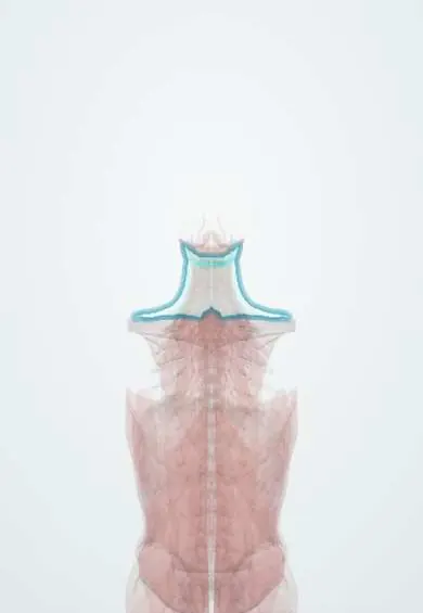
Platysma !
The platysma is a thin sheet of muscle just under the skin of the front of the neck; it tightens the neck skin.
Opens in the Durgical app, no sign-up needed.
What it is
A thin sheet of muscle just under the skin of the front of the neck.
What it does
Tightens the neck skin. It is the first layer opened in an ACDF incision.
Key facts
- Origin
- Fascia over upper pectoralis major and deltoid
- Insertion
- Inferior border of the mandible, skin of lower face and angle of the mouth
- Action
- Tenses the skin of the neck; depresses mandible and corners of the mouth
- Nerve
- Cervical branch of the facial nerve (CN VII)
- Blood supply
- Submental (from facial) and suprascapular arteries
Relations
- Superficial
- Subcutaneous fat and the skin of the neck
- Deep
- Investing layer of deep cervical fascia; external and anterior jugular veins; great auricular, transverse cervical and supraclavicular nerves; sternocleidomastoid
- Above
- Lower border of the mandible and the muscles at the corner of the mouth; the marginal mandibular branch of the facial nerve near the jaw
- Below
- Fascia over pectoralis major and deltoid, below the clavicle
- Medial
- The two muscles leave a midline gap in the lower neck and often interlace below the chin
How to find it
Clenching the teeth and pulling the corners of the mouth down and out (a grimace) tightens the skin of the neck, showing vertical ridges from the jaw to the clavicle.
Examination
- Facial nerve exam: asked to grimace, a platysma that does not tense on one side fits a lesion of the facial nerve or its cervical branch
On imaging
Ultrasound, CT and MRI show it as a thin layer under the skin of the front of the neck, with the external jugular vein beneath it. It is a landmark rather than a target: a lump superficial to it lies in the skin or fat, while one deep to it lies in the deeper neck. After neck surgery, ultrasound or CT shows a collection or haematoma in the subplatysmal plane.
Clinical notes
- Subplatysmal flaps are the standard plane in thyroid, carotid and anterior spine surgery
- The vertical platysmal bands of the ageing neck appear when its medial edges separate and become slack
Anatomical variants
- How far its fibres reach (up into the face, down onto the chest, across the midline) varies between people
Development
A muscle of facial expression that comes from the second (hyoid) pharyngeal arch; it spreads down into the neck carrying a branch of the facial nerve, which is why it is supplied by the cervical branch of CN VII rather than by cervical spinal nerves.
Test yourself
Think of the answer, then tap the question to see it.
What nerve supplies the platysma?
The cervical branch of the facial nerve (CN VII).
Why are neck flaps raised in the subplatysmal plane?
It protects the marginal mandibular branch of the facial nerve, which lies deep to the platysma near the jaw.
How is the platysma tested at the bedside?
Asking the person to grimace: a platysma that does not tense on one side fits a lesion of the facial nerve or its cervical branch.
Educational information, not medical advice. For any symptom, see a health professional.
Its names
- Official Latin (TA2)
- platysma TA2 2147
- Official Spanish (DeCS)
- Platisma
- English (TA2)
- Platysma
- Also called
- platysma muscle · musculus platysma
Related
Sources: Terminologia Anatomica (TA2); Anatomy, Head and Neck, Platysma; Anatomy of the Human Body (20th ed.): Development of the Muscles (Lea & Febiger (Gray's Anatomy, 1918), 1918) · adapted, Public domain.
Spotted a mistake?We are always improving for the community. Tell us what is wrong and our team will review it.Report a mistake
Representative model, for educational purposes only.