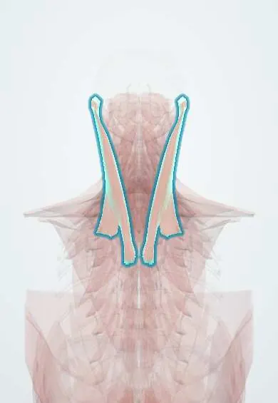
Sternocleidomastoid !
The sternocleidomastoid is the large strap muscle on each side of the neck, from the breastbone and collarbone to behind the ear; it turns the head.
Official name: Sternocleidomastoid muscle
Opens in the Durgical app, no sign-up needed.
What it is
The large strap muscle on each side of the neck, from the breastbone and collarbone to behind the ear.
What it does
Turns the head to the opposite side and tilts it to the same side; both together bend the neck forward. In an ACDF the incision follows its front edge.
Key facts
- Origin
- Sternal head: anterior manubrium; clavicular head: medial third of clavicle
- Insertion
- Mastoid process and lateral half of the superior nuchal line
- Action
- Unilateral: tilts head to same side, turns face to opposite side; bilateral: flexes neck; accessory inspiration
- Nerve
- Spinal accessory nerve (CN XI); proprioception C2–C3
- Blood supply
- Occipital and superior thyroid arteries (sternocleidomastoid branches)
Relations
- Superficial
- Platysma, the external jugular vein crossing it obliquely, and the great auricular and transverse cervical nerves
- Deep
- Carotid sheath (common and internal carotid arteries, internal jugular vein, vagus nerve), the cervical plexus, the scalenes and, above, splenius capitis
- In front
- Anterior triangle of the neck, with its submandibular, submental, carotid and muscular triangles
- Behind
- Posterior triangle: the spinal accessory nerve crossing it, the brachial plexus trunks low down and the inferior belly of omohyoid
- Below
- The small hollow between its sternal and clavicular heads lies over the internal jugular vein; this is the lesser supraclavicular fossa (Sédillot triangle), the landmark for the central approach to the internal jugular vein
How to find it
Turning the head to the opposite side against resistance makes the muscle stand out from behind the ear to the sternoclavicular joint; the two heads and the small hollow between them are seen just above the clavicle.
Examination
- Head turn against resistance tests the accessory nerve: weakness turning the face away from a side points to the nerve on that side
- Weakness of both sternocleidomastoid and trapezius points to a CN XI lesion high up (skull base, jugular foramen); trapezius alone points to the posterior triangle
On imaging
Ultrasound is the first test in an infant with torticollis: the muscle is thickened, sometimes with a focal mass inside it (fibromatosis colli) and normal tissue around it, which usually avoids a biopsy. On neck CT or MRI it is the landmark between the anterior and posterior triangles; the carotid sheath and the level II–IV lymph nodes lie deep to it.
Clinical notes
- Congenital muscular torticollis: a fibrotic, shortened muscle, sometimes with a lump in infancy, tilts the head to the same side and turns the chin to the opposite side
- Its anterior border guides incisions for anterior cervical spine surgery, carotid endarterectomy and neck dissection
- The hollow between its two heads is a landmark for internal jugular vein catheterisation
- Often involved in cervical dystonia (spasmodic torticollis), where it is a common target for botulinum toxin injections
Anatomical variants
- Rarely absent; its origins vary, especially the width of the clavicular head
Development
It and trapezius come from a single muscle mass at the back of the developing head, just behind the last pharyngeal arch. The accessory nerve grows into this mass early and is carried down as it spreads toward the shoulder girdle, so both muscles share CN XI; whether the mass is pharyngeal-arch or somite mesoderm is still debated.
Related conditions
- Whiplash (cervical sprain)
- Torticollis
- Myotonic dystrophy (types 1 and 2)
- Dystonia (generalised and focal)
- Thyroglossal duct cyst and branchial cleft cyst
Test yourself
Think of the answer, then tap the question to see it.
What nerve supplies the sternocleidomastoid?
The spinal accessory nerve (CN XI); C2–C3 carry proprioception.
What does the sternocleidomastoid do when it contracts on one side?
It tilts the head to the same side and turns the face to the opposite side; together both flex the neck.
What is the nerve point of the neck on the sternocleidomastoid?
The middle of its posterior border, where the cutaneous cervical plexus nerves wind round.
What does weakness of both sternocleidomastoid and trapezius suggest?
A high CN XI lesion (skull base, jugular foramen); trapezius alone points to the posterior triangle.
What is congenital muscular torticollis of the sternocleidomastoid?
A fibrotic, shortened muscle that tilts the head to the same side and turns the chin to the opposite side.
Educational information, not medical advice. For any symptom, see a health professional.
Its names
- Official Latin (TA2)
- musculus sternocleidomastoideus TA2 2156
- Official Spanish (DeCS)
- Músculo esternocleidomastoideo DeCS D009334
- English (TA2)
- Sternocleidomastoid muscle
- Also called
- sternomastoid muscle
Related
Sources: Terminologia Anatomica (TA2); DeCS; Anatomy, Head and Neck: Sternocleidomastoid Muscle; Anatomy of the Human Body (20th ed.): Development of the Muscles (Lea & Febiger (Gray's Anatomy, 1918), 1918) · adapted, Public domain; Anatomía humana (4.ª ed.), tomo 1, cap. 19: Músculos del cuello.
Spotted a mistake?We are always improving for the community. Tell us what is wrong and our team will review it.Report a mistake
Representative model, for educational purposes only.