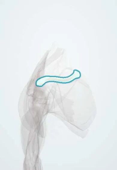
Clavicle !
The clavicle, or collarbone, is the long bone at the front of the shoulder; it holds the arm out to the side and passes its forces to the trunk.
Opens in the Durgical app, no sign-up needed.
What it is
The S-shaped collarbone between the breastbone and the shoulder blade.
What it does
Holds the shoulder out to the side and passes arm forces to the trunk.
Key facts
- Joints with
- Sternoclavicular joint (manubrium, 1st costal cartilage); acromioclavicular joint (acromion)
- Landmarks
- Medial 2/3 convex forward, lateral 1/3 concave; conoid tubercle, trapezoid line; fractures most often at middle–lateral third junction
Relations
- Above
- Clavicular head of the sternocleidomastoid medially and the upper trapezius laterally; the supraclavicular nerves cross it under the skin and are at risk in plate fixation
- In front
- Origin of the clavicular head of the pectoralis major medially and of the front deltoid laterally
- Below
- Subclavius muscle in its groove; conoid tubercle and trapezoid line with the coracoclavicular ligaments; the costoclavicular ligament ties the medial end to the first rib
- Behind
- Subclavian vein, subclavian artery and the divisions of the brachial plexus run behind the medial two thirds through the costoclavicular space
- Medial
- Sternoclavicular joint with the manubrium and the SC disc, held mainly by the SC ligaments and the costoclavicular ligament
- Lateral
- Acromioclavicular joint with the acromion, with the AC disc and the AC ligament
How to find it
Subcutaneous along its whole length: it is followed from the jugular notch outward along its S-curve to the acromioclavicular joint, felt as a small step just medial to the acromion. A fracture shows as a visible bump or tenting of the skin over the shaft.
On imaging
An AP view plus a second view with the beam tilted toward the head shows shaft fractures and their shortening. The acromioclavicular joint needs a softer exposure with the beam tilted slightly toward the head (Zanca view). The sternoclavicular joint is poorly seen on plain films: CT is the standard for medial-end injuries.
Common problems
- Fracture, usually in the middle third, from a fall on the shoulder or an outstretched hand.
- Wear or arthritis at either end (AC or SC joint).
How it is diagnosed
Exam for a bump, tenderness and skin tenting. X-rays show the fracture and how far the ends have shifted.
Treatment
- Most heal in a sling with early gentle movement.
- Surgery (plate and screws or a rod) when the ends are badly shifted or shortened, or the skin is at risk.
Clinical notes
- Lateral-third fractures that leave the medial fragment detached from the coracoclavicular ligaments are unstable and heal less reliably than midshaft fractures
- Posterior sternoclavicular dislocation can press on the trachea, oesophagus and great vessels behind the manubrium: a medical emergency
- The most frequently fractured bone during birth; the newborn moves the arm less and the Moro reflex is weaker on that side
- A bulky callus or malunion can narrow the costoclavicular space and compress the brachial plexus or subclavian vessels
Anatomical variants
- Sex differences: in men it tends to be longer, thicker and more strongly curved
- A deep rhomboid fossa (impression of the costoclavicular ligament) under the medial end can look like an erosion on X-ray
Development
The first bone in the body to start ossifying (around the fifth to sixth fetal week), mostly by intramembranous ossification. Its medial epiphysis is the last of the body to fuse, in the early to mid-twenties, so in young adults a physeal fracture can imitate a sternoclavicular dislocation. Clavicles are underdeveloped or absent in cleidocranial dysplasia.
Related conditions
Test yourself
Think of the answer, then tap the question to see it.
In a midshaft clavicle fracture, which muscles displace the two fragments?
The sternocleidomastoid pulls the medial fragment up; the weight of the arm drops the lateral fragment and the pectoralis major draws it medially, shortening the shoulder.
Which neurovascular structures pass behind the medial two thirds of the clavicle?
The subclavian vein, subclavian artery and brachial plexus, crossing the first rib through the costoclavicular space; the subclavius lies between them and the bone.
Which ligaments hang the scapula from the clavicle, and why does their state matter in acromioclavicular separations?
The coracoclavicular ligaments (conoid and trapezoid); whether they are torn separates mild from severe acromioclavicular separations.
Why is a posterior sternoclavicular dislocation of the clavicle a medical emergency?
The medial clavicle can press on the trachea, oesophagus and great vessels behind the manubrium.
Which imaging study is the standard for injuries of the medial end of the clavicle?
CT, because the sternoclavicular joint is poorly seen on plain films.
Educational information, not medical advice. For any symptom, see a health professional.
Its names
- Official Latin (TA2)
- clavicula TA2 1168
- Official Spanish (DeCS)
- Clavícula
- English (TA2)
- Clavicle
- Also called
- collarbone
Related
- AC capsule
- AC disc
- AC ligament
- Conoid ligament
- Costoclavicular ligament
- Front deltoid
- Interclavicular ligament
- Pectoralis major
Sources: Terminologia Anatomica (TA2); Anatomy, Shoulder and Upper Limb, Clavicle.
Spotted a mistake?We are always improving for the community. Tell us what is wrong and our team will review it.Report a mistake
Representative model, for educational purposes only.