Muscular system · Head and neck
Cervical fascia !
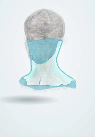
The cervical fascia is a set of tough sheets that wrap the muscles and organs of the neck into compartments, which can limit or guide infections.
Official name: Superficial investing cervical fascia
Opens in the Durgical app, no sign-up needed.
What it is
A sleeve of fascia wrapping the neck and splitting around the SCM and trapezius.
What it does
Divides the neck into compartments that limit (or guide) the spread of infection.
Key facts
- Attaches
- Investing layer: superior nuchal line, mastoid, mandible and hyoid above; clavicle, manubrium and acromion below
- Function
- Encloses the sternocleidomastoid and trapezius; with the pretracheal and prevertebral layers it makes the neck compartments that guide infection
Relations
- Contents
- Investing layer: sternocleidomastoid, trapezius, submandibular and parotid glands; pretracheal layer: thyroid gland, parathyroid glands, larynx, trachea, pharynx, oesophagus and strap muscles; prevertebral layer: deep neck muscles, phrenic nerve and brachial plexus roots
- Superficial
- Superficial fascia with the platysma, the external jugular vein and the cutaneous nerves of the cervical plexus
- Deep
- The prevertebral layer lies on the vertebral column and the deep neck muscles; the sympathetic trunk runs on it
How to find it
Not palpable. In surgery the investing layer is the first tough sheet under the platysma, opened along the front of the sternocleidomastoid; above the manubrium it splits to form the suprasternal space, which holds the jugular venous arch.
On imaging
Contrast CT is the standard way to map a deep neck infection: it shows which space is involved (parapharyngeal, retropharyngeal, danger, prevertebral, submandibular) and any spread into the mediastinum.
Clinical notes
- Infection behind the prevertebral layer, such as tuberculosis of the cervical spine, can track down to the mediastinum or along the axillary sheath into the axilla
- A haematoma after thyroid surgery is trapped under the strap muscles and pretracheal layer and can compress the airway; opening the wound urgently is the taught emergency step
- Neck wounds that breach the platysma are classed as penetrating neck injuries
Related conditions
Test yourself
Think of the answer, then tap the question to see it.
What are the three layers of the deep cervical fascia?
Investing, pretracheal and prevertebral; the carotid sheath takes a contribution from all three.
Which space lets a retropharyngeal infection reach the posterior mediastinum?
The danger space, between the alar and prevertebral layers, running from the skull base to the diaphragm: descending necrotizing mediastinitis.
Why does the thyroid gland move on swallowing?
The pretracheal layer of the cervical fascia wraps the thyroid and ties it to the larynx.
Why is a haematoma after thyroid surgery an emergency?
It is trapped under the strap muscles and pretracheal layer and can compress the airway; opening the wound urgently is the taught step.
Educational information, not medical advice. For any symptom, see a health professional.
Its names
- Official Latin (TA2)
- fascia investiens superficialis colli TA2 2207
- Official Spanish (DeCS)
- Lámina superficial de la fascia cervical DeCS D005205
- English (TA2)
- Superficial investing cervical fascia
- Also called
- investing layer of cervical fascia · superficial layer of deep cervical fascia · fascia investiens colli · fascia superficialis colli
Sources: Terminologia Anatomica (TA2); DeCS; Anatomy, Head and Neck, Deep Cervical Neck Fascia; Anatomy, Head and Neck: Carotid Sheath.
Spotted a mistake?We are always improving for the community. Tell us what is wrong and our team will review it.Report a mistake
Representative model, for educational purposes only.