Cardiovascular system · Head and neck
External jugular vein !
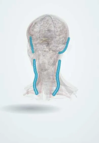
The external jugular vein runs down the side of the neck just under the skin, draining the scalp and face; it bulges when chest pressure rises.
Opens in the Durgical app, no sign-up needed.
What it is
A surface vein crossing the sternocleidomastoid, visible when you strain.
What it does
Drains the scalp and face; it bulges when pressure in the chest is high.
Key facts
- Drains
- Scalp and face (posterior retromandibular + posterior auricular veins)
- Into
- Subclavian vein
Relations
- Course
- Begins near the angle of the mandible, below the parotid gland, runs down and back across the sternocleidomastoid, enters the posterior triangle above the clavicle, pierces the deep fascia and ends in the subclavian vein
- Superficial
- Platysma and skin
- Deep
- Sternocleidomastoid; lower down, the investing layer of deep cervical fascia roofing the posterior triangle
- Behind
- The great auricular nerve, which climbs across the sternocleidomastoid roughly parallel to it
How to find it
With the person lying flat or slightly head-down and the head turned to the other side, or during a brief strain (Valsalva), it fills and is seen running from the angle of the jaw toward the middle of the clavicle.
On imaging
On ultrasound it is a superficial, easily compressed vein under the platysma, crossing the sternocleidomastoid; ultrasound guides cannulation when it cannot be seen. After a catheter, thrombosis shows as a vein that does not compress, with echogenic material and no Doppler flow. On contrast CT it is followed down to the subclavian vein. Its filling is a poor guide to central venous pressure.
Clinical notes
- Cannulation: a head-down tilt distends it; the angle at which it crosses the clavicle and its valves near the lower end can make it hard to advance a catheter
- Distended when right-sided pressure is high (heart failure, cardiac tamponade, tension pneumothorax), but the valves and fascia make it less reliable than the internal jugular for estimating pressure
- Often tied off in neck dissection and parotid surgery without harm, thanks to rich venous collaterals
Anatomical variants
- Its size varies widely; it may be duplicated, small or absent, and it sometimes ends in the internal jugular vein instead of the subclavian vein
Development
At first it drains only the area behind the ear; cross-links with the embryonic linguofacial vein later hand it the posterior veins of the face, which explains why its size and ending vary so much.
Test yourself
Think of the answer, then tap the question to see it.
What is the course of the external jugular vein in the neck?
From the angle of the mandible obliquely across the sternocleidomastoid under the platysma, to the subclavian vein above the middle of the clavicle.
Why can a cut external jugular vein above the clavicle cause an air embolism?
Where it pierces the deep cervical fascia, the fascia holds the cut vein open so it can suck in air.
Why is the internal jugular vein preferred over the external jugular vein to estimate venous pressure?
The valves and fascia of the external jugular vein make its filling only a rough pressure guide.
Educational information, not medical advice. For any symptom, see a health professional.
Its names
- Official Latin (TA2)
- vena jugularis externa TA2 4956
- Official Spanish (DeCS)
- Vena yugular externa DeCS D007601
- English (TA2)
- External jugular vein
- Also called
- vena jugularis superficialis dorsalis
Sources: Terminologia Anatomica (TA2); DeCS; Anatomy, Head and Neck, External Jugular Veins; Anatomy of the Human Body (20th ed.): Development of the Vascular System (Lea & Febiger (Gray's Anatomy, 1918), 1918) · adapted, Public domain.
Spotted a mistake?We are always improving for the community. Tell us what is wrong and our team will review it.Report a mistake
Representative model, for educational purposes only.