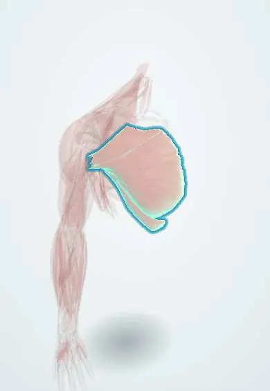
Pectoralis major !
The pectoralis major is the large fan-shaped chest muscle; it pulls the arm across the body, turns it inward and powers pushing.
Opens in the Durgical app, no sign-up needed.
What it is
The large fan-shaped chest muscle from the collarbone and breastbone to the humerus.
What it does
Pulls the arm across the body and turns it inward; powers pushing.
Key facts
- Origin
- Clavicular head: medial half of clavicle; sternocostal head: sternum, costal cartilages 1–6, external oblique aponeurosis
- Insertion
- Lateral lip of intertubercular sulcus (crest of greater tubercle) of humerus
- Action
- Adducts and medially rotates humerus; clavicular head flexes, sternocostal head extends from flexion
- Nerve
- Lateral pectoral nerve (C5–C7) and medial pectoral nerve (C8–T1)
- Blood supply
- Pectoral branch of thoracoacromial artery; perforating branches of internal thoracic artery
Relations
- Superficial
- The breast on the pectoral fascia, separated by the loose retromammary space; skin
- Deep
- Pectoralis minor, subclavius and the clavipectoral fascia; lower down the ribs, intercostal muscles and serratus anterior
- Lateral
- Front deltoid across the deltopectoral groove, with the cephalic vein
- Above
- The clavicle; the clavipectoral (deltopectoral) triangle opens between the clavicle, its clavicular head and the deltoid
- Below
- Its free lower border, the anterior axillary fold, overlying the axillary lymph nodes
How to find it
Pressing the hands together in front of the chest contracts the whole muscle. Resisted horizontal adduction with the arm forward at 90° brings out the clavicular head; resisted adduction down toward the opposite hip brings out the sternocostal head. The anterior axillary fold can be gripped between thumb and fingers.
Examination
- Clavicular head (lateral pectoral nerve): resisted horizontal adduction from 90° of flexion; sternocostal head (medial and lateral pectoral nerves): resisted adduction from abduction; isolated weakness of one head localises a pectoral nerve or root lesion
On imaging
MRI is best for tears: axial and oblique coronal images follow the tendon to its insertion lateral to the bicipital groove; the classic injury is avulsion of the lower sternal fibres while bench pressing. Ultrasound is a quick first check of tendon continuity and haematoma. On a chest radiograph, its absence (Poland syndrome) or a mastectomy makes that side look darker.
Common problems
- Tendon rupture, usually during a heavy bench press: a pop, bruising and a changed chest contour.
- Muscle strains.
How it is diagnosed
Exam of the armpit fold and pushing strength. MRI shows where and how much it is torn.
Treatment
- Surgical repair of the tendon to the humerus for active people.
- Without surgery for partial tears and muscle strains.
Clinical notes
- Pedicled pectoralis major flap on the pectoral branch of the thoracoacromial artery: a classic reconstruction for head and neck defects
- Breast examination with the hands pressed on the hips tenses the muscle; a lump that becomes fixed suggests a tumour attached to the pectoral fascia or muscle
- The pectoralis major tendon transfer is used for irreparable subscapularis tears
Anatomical variants
- Heads may be more or less separate and attachments vary; the sternocostal head may be missing (Poland syndrome, sometimes with hand anomalies on the same side)
- A sternalis muscle, a vertical strip over the sternum, is a related variant that can look like a mass on mammography
Development
It begins next to the arm bud at lower cervical levels and migrates over the front of the chest, bringing its brachial plexus nerves with it; failure of the sternocostal head to form is the basis of Poland syndrome.
Memory aid
The medial pectoral nerve lies lateral to the lateral pectoral nerve: they are named for their cords, not their position.
Related conditions
Test yourself
Think of the answer, then tap the question to see it.
What is the nerve supply of the two heads of the pectoralis major?
Clavicular head: lateral pectoral nerve; sternocostal head: medial and lateral pectoral nerves.
Which structure runs in the deltopectoral groove, and where does it go?
The cephalic vein, up to the clavipectoral triangle, where it pierces the clavipectoral fascia.
Where does the pectoralis major insert, and what are its main actions?
On the lateral lip of the intertubercular sulcus; it adducts and medially rotates the humerus.
Absence of which part of the pectoralis major is the core of Poland syndrome?
The sternocostal head.
What vessel does a pedicled pectoralis major flap rely on?
The pectoral branch of the thoracoacromial artery; a classic flap for head and neck defects.
Educational information, not medical advice. For any symptom, see a health professional.
Its names
- Official Latin (TA2)
- musculus pectoralis major TA2 2301
- Official Spanish (DeCS)
- Músculo pectoral mayor
- English (TA2)
- Pectoralis major
Related
- Cephalic vein
- Interpectoral nodes
- Pectoral nerves
- Thoracoacromial artery
- Lateral thoracic artery
- Clavicle
- Manubrium
- Pec major tendon
Sources: Terminologia Anatomica (TA2); Anatomy, Shoulder and Upper Limb, Pectoral Muscles; Anatomy of the Human Body (20th ed.): Development of the Muscles (Lea & Febiger (Gray's Anatomy, 1918), 1918) · adapted, Public domain.
Spotted a mistake?We are always improving for the community. Tell us what is wrong and our team will review it.Report a mistake
Representative model, for educational purposes only.