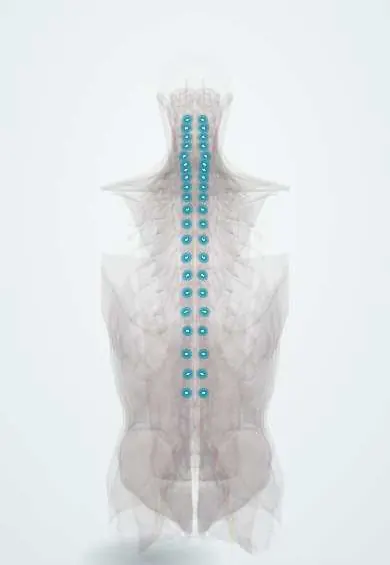
Dorsal root ganglia !
The dorsal root ganglia are small swellings on the sensory nerve roots, in the side openings of the spine; they relay all body sensation.
Official name: Spinal ganglion
Opens in the Durgical app, no sign-up needed.
What it is
Small swellings on the sensory roots, in each side opening of the spine, holding the cell bodies of the sensory nerves.
What it does
Relay all sensation from the body to the cord. Very sensitive to pressure: one reason a pinched root hurts so much.
Key facts
- Where
- On each dorsal root in the intervertebral foramen, just outside the dural sac (sacral ganglia inside the sacral canal)
- Function
- Cell bodies of pseudounipolar primary sensory neurons; no synapses
Relations
- Above
- The pedicle of the vertebra above, under which it sits in the foramen
- In front
- The disc and vertebral body (in the neck, the uncovertebral joint); the ventral root runs in front of it
- Behind
- The facet joint, formed here by the superior articular process of the vertebra below
- Lateral
- Just beyond it the dorsal and ventral roots join into the spinal nerve, which splits into dorsal and ventral rami; in the neck the vertebral artery lies in front of the nerve
How to find it
It cannot be felt. Most lie in the intervertebral foramen, just under the pedicle and outside the dural sac; the exceptions are the upper two cervical ganglia, which sit beside the arches of the atlas and axis, and the sacral and coccygeal ones, which stay inside the sacral canal.
On imaging
On MRI it is an oval swelling in the foramen that normally enhances with contrast (it has no blood-nerve barrier), which should not be mistaken for a tumour. Perineural (Tarlov) cysts on the sacral roots are common incidental findings.
Common problems
- Cervical radiculopathy: a disc or bone spur pinches a root, causing pain, numbness or weakness down the arm in that root's pattern.
How it is diagnosed
Exam of strength, reflexes and feeling in the arm (each root has its own pattern); MRI; sometimes nerve tests (EMG).
Treatment
- Most improve with time, physical therapy, medicine and sometimes an injection.
- Lasting pain or weakness: surgery to free the root, such as ACDF or a foraminotomy.
Clinical notes
- Postherpetic neuralgia: pain that persists after shingles has healed, from damage in the ganglion and its nerve
- Sensory neuronopathy (ganglionopathy) from Sjögren syndrome, paraneoplastic disease or toxins: patchy, non-length-dependent sensory loss and sensory ataxia
- Target of transforaminal injections and pulsed radiofrequency for chronic radicular pain
Development
The ganglia come from neural crest cells, like the autonomic ganglia, which is why they lie outside the neural tube.
Related conditions
Test yourself
Think of the answer, then tap the question to see it.
Where are the dorsal root ganglia, and what do they contain?
On each dorsal root in the intervertebral foramen, just outside the dural sac; they hold the cell bodies of pseudounipolar sensory neurons, with no synapses.
How do sensory nerve action potentials separate a root avulsion from a more distal lesion?
A lesion proximal to the dorsal root ganglion leaves the sensory potential normal in a numb limb; a distal lesion abolishes it.
Which virus lies dormant in the dorsal root ganglia?
Varicella-zoster virus, which reactivates as shingles in that dermatome, most often a thoracic one.
Educational information, not medical advice. For any symptom, see a health professional.
Its names
- Official Latin (TA2)
- ganglion spinale TA2 6167
- Official Spanish (DeCS)
- Ganglios espinales DeCS D005727
- English (TA2)
- Spinal ganglion
- Also called
- sensory ganglion of spinal nerve · dorsal root ganglion · Ganglia, Spinal · ganglion spinale sensoriale
Related
Sources: Terminologia Anatomica (TA2); DeCS; Neuroanatomy, Spinal Nerves.
Spotted a mistake?We are always improving for the community. Tell us what is wrong and our team will review it.Report a mistake
Representative model, for educational purposes only.