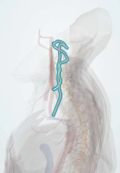
Vertebral artery !
The vertebral artery runs up each side of the neck through holes in C6 to C1, then into the skull; it feeds the back of the brain.
Opens in the Durgical app, no sign-up needed.
What it is
An artery on each side of the neck that runs up through holes in the transverse processes of C6 to C1, then into the skull.
What it does
Supplies the back of the brain, the brainstem and the top of the spinal cord.
Key facts
- Origin
- First part of the subclavian artery; ascends through transverse foramina C6–C1; enters skull via foramen magnum
- Branches
- Spinal (radicular) and muscular branches in the neck; anterior and posterior spinal; PICA; joins the other to form the basilar artery
- Supplies
- Cervical cord, deep neck muscles, brainstem, cerebellum, occipital lobes (via basilar)
Relations
- Course
- Rises from the subclavian artery between the longus colli and scalenus anterior, enters the C6 transverse foramen, climbs through the foramina to the atlas, curves back behind its lateral mass in a groove on the posterior arch, pierces the atlanto-occipital membrane and dura and enters the skull through the foramen magnum
- In front
- At the root of the neck, the common carotid artery and the vertebral vein in front of its first part, with the inferior thyroid artery arching across it at about C7 (on the left, also the thoracic duct)
- Medial
- Uncovertebral joints and vertebral bodies; osteophytes from these joints can press on it
- Behind
- First part: the C7 transverse process, the cervicothoracic (stellate) ganglion and the ventral rami of C7 and C8; higher up, the ventral rami of the cervical spinal nerves, which pass behind it as they leave the foramina
- Above
- At the atlas it lies in the suboccipital triangle, with the suboccipital nerve between it and the posterior arch
How to find it
It cannot be felt. In posterior exposures it lies in the suboccipital triangle (rectus capitis posterior major and the two obliques) on the posterior arch of the atlas; in anterior surgery it lies just lateral to the uncovertebral joints, which mark the safe lateral limit.
On imaging
CT or MR angiography shows its course and dominance (the left is often larger); axial CT shows whether a fracture enters a transverse foramen.
Common problems
- Can be torn (dissection) in neck trauma or, rarely, forceful neck manipulation: a cause of stroke in younger people.
- Sets the side limit in anterior neck surgery and the path for screws at C1-C2.
How it is diagnosed
CT or MR angiography.
Treatment
- Dissections are usually treated with blood thinners; surgeons study its course on scans before neck surgery.
Clinical notes
- In posterior C1 exposure, dissection along the upper border of the posterior arch is usually kept within about 1.5 cm of the midline to avoid the artery
- C1–C2 transarticular screws and C2 pedicle or pars screws need its path checked on CT first, as a high-riding artery can make them unsafe
- Rotational vertebral artery occlusion (bow hunter's syndrome): turning the head compresses the artery near C1–C2, causing dizziness or fainting
Anatomical variants
- The left artery arises directly from the aortic arch in about 1 in 20 people
- It enters the C6 foramen in most people (about 9 in 10), otherwise at C5, C4 or C7
- A bony bridge over the groove on the atlas (ponticulus posticus) can be mistaken for a wide posterior arch when C1 screws are placed
Development
Its neck part forms from a longitudinal chain linking the upper cervical segmental arteries, and the seventh segmental artery becomes its origin from the subclavian; this explains entry into a foramen other than C6 and origins from the aortic arch.
Related conditions
- Transient ischaemic attack
- Brainstem stroke syndromes (Wallenberg and others)
- Subclavian steal syndrome
Test yourself
Think of the answer, then tap the question to see it.
Where does the vertebral artery arise, and at which level does it usually enter the transverse foramina?
From the first part of the subclavian artery; it usually enters the C6 transverse foramen (about 9 in 10 people) and climbs to C1.
What are the four segments of the vertebral artery?
V1 subclavian to C6; V2 in the transverse foramina C6–C2; V3 looping behind the atlas; V4 inside the skull to the basilar artery.
In which triangle does the vertebral artery lie on the posterior arch of the atlas?
The suboccipital triangle, with the suboccipital nerve between the artery and the posterior arch.
What is subclavian steal, and how does it involve the vertebral artery?
A blocked proximal subclavian artery draws reversed flow down the vertebral artery, so arm exercise can cause dizziness.
Which landmark marks the safe lateral limit for the vertebral artery in anterior cervical surgery?
The uncovertebral joints: the vertebral artery lies just lateral to them.
Educational information, not medical advice. For any symptom, see a health professional.
Its names
- Official Latin (TA2)
- arteria vertebralis TA2 4538
- Official Spanish (DeCS)
- Arteria vertebral DeCS D014711
- English (TA2)
- Vertebral artery
Related
- Vertebral vein
- Anterior spinal artery
- Suboccipital muscles
- Atlas (C1)
- Axis (C2)
- C3 vertebra
- C4 vertebra
- C5 vertebra
Sources: Terminologia Anatomica (TA2); DeCS; Anatomy, Head and Neck: Cervical Vertebrae; Anatomy, Head and Neck, Occipital Bone, Artery, Vein, and Nerve; Anatomy of the Human Body (20th ed.): Development of the Vascular System (Lea & Febiger (Gray's Anatomy, 1918), 1918) · adapted, Public domain; Tratado de anatomía humana (9.ª ed.), tomo II, Libro IV Angiología, arterias subclavia, axilar, humeral, radial y cubital, arcos palmares y vías anastomóticas del miembro superior.
Spotted a mistake?We are always improving for the community. Tell us what is wrong and our team will review it.Report a mistake
Representative model, for educational purposes only.