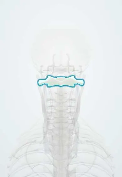
Atlas (C1) !
The atlas (C1) is the first neck vertebra, a ring with no body that carries the skull; it lets the head nod and turns on C2.
Official name: Atlas
Opens in the Durgical app, no sign-up needed.
What it is
The first neck vertebra, a ring with no body that carries the skull.
What it does
Lets you nod 'yes' where it meets the skull, and turns on the axis (C2) for about half of all neck rotation.
Key facts
- Joints with
- Occipital condyles above (atlanto-occipital joints); axis below: dens and C2 superior facets (median and lateral atlantoaxial joints)
- Landmarks
- No body and no spinous process; anterior and posterior arches; lateral masses; facet for dens on anterior arch; groove for vertebral artery on posterior arch; wide transverse processes
Relations
- Above
- The atlanto-occipital membranes close the gaps up to the base of the skull; the vertebral artery pierces the posterior membrane to reach the foramen magnum
- In front
- Longus colli attaches to the anterior tubercle; in front lie the prevertebral fascia, the retropharyngeal space and the back wall of the pharynx
- Behind
- Suboccipital muscles: rectus capitis posterior minor arises from the posterior tubercle; the posterior arch forms the floor of the suboccipital triangle, with the vertebral artery and the suboccipital nerve
- Lateral
- The wide transverse process lies between the mastoid process and the angle of the mandible; the accessory nerve crosses it and the internal jugular vein lies just in front
- Contents
- Behind the dens, kept apart from it by the transverse ligament of the atlas: the spinal cord passing into the medulla, its meninges and the tectorial membrane
How to find it
Mostly too deep to feel. With firm, gentle pressure the tip of the transverse process is felt as a hard point in the hollow between the mastoid process and the angle of the mandible. There is no spinous process, so the first midline bony point felt below the occiput belongs to the axis (C2).
Examination
- Sharp–Purser test (suspected atlantoaxial instability, as in rheumatoid arthritis; never after acute trauma): seated with the neck slightly bent, the C2 spine is steadied and the forehead pushed gently backward; a slide or clunk with easing of symptoms suggests C1 had slipped forward on C2
On imaging
Open-mouth AP view: the outer edges of the lateral masses should line up with those of C2, and the gaps either side of the dens should match (head rotation can make them unequal). Lateral view: the atlantodens interval, commonly quoted as up to about 3 mm in adults and 5 mm in children. CT with reconstructions shows ring fractures, which usually break in more than one place.
Common problems
- Fractures from a heavy blow to the top of the head, such as diving into shallow water (Jefferson fracture).
- Instability between C1 and C2 if the transverse ligament is torn or weakened, for example by rheumatoid arthritis.
How it is diagnosed
CT shows fractures of the ring best. Flexion-extension X-rays or MRI check the ligaments and whether C1 slides on C2.
Treatment
- Stable fractures: a rigid collar or halo brace for several weeks.
- Unstable injuries: surgery to fuse C1 to C2 (or the skull to C2) with screws, rods and bone graft.
Clinical notes
- Isolated posterior arch fractures come from hyperextension and are usually stable; C1 fractures often come with C2 fractures, so the whole cervical spine is imaged
- Atlantoaxial rotatory subluxation in children, sometimes after a throat infection (Grisel syndrome) or head and neck surgery, shows as a fixed head tilt and turn (torticollis)
- Ligament laxity in Down syndrome lets C1 slide forward on C2, a recognised risk during anesthesia and contact sports
- Posterior C1–C2 fusion uses screws in the C1 lateral masses: the C2 nerve root and a large venous plexus lie just below the posterior arch, over the lateral atlantoaxial joint
Anatomical variants
- Ponticulus posticus (arcuate foramen): a bony bridge that turns the vertebral artery groove into a tunnel; visible on lateral X-rays and relevant when placing screws
- Occipitalization (assimilation) of the atlas: partial or complete fusion with the base of the skull, sometimes with basilar invagination or a Chiari malformation
- Clefts or partial absence of the arches, most often a midline gap in the posterior arch, which can mimic a fracture
Development
Usually three ossification centres: one for the anterior arch and one for each lateral mass with its half of the posterior arch. The anterior arch may still be cartilage at birth; the gaps (synchondroses) close in early childhood and should not be read as a fracture.
Memory aid
Steel's rule of thirds: inside the ring of the atlas, one third is dens, one third spinal cord and one third free space, which is why some C1 slip is tolerated before the cord is squeezed.
Related conditions
Test yourself
Think of the answer, then tap the question to see it.
What mechanism causes a Jefferson fracture of the atlas (C1), and why is the spinal cord often spared?
An axial load on the top of the head, as in diving into shallow water: the ring splits in front and behind and the lateral masses spread, so the canal widens.
On the open-mouth view, what does overhang of the lateral masses of the atlas (C1) on C2 totalling more than about 7 mm suggest?
A torn transverse ligament of the atlas and an unstable injury (rule of Spence).
Which artery runs in a groove on the upper surface of the posterior arch of the atlas (C1)?
The vertebral artery, with the C1 spinal nerve; this is why posterior exposure of C1 stays close to the midline.
According to Steel's rule of thirds, what fills the ring of the atlas (C1)?
One third dens, one third spinal cord and one third free space, so some C1 slip is tolerated before the cord is squeezed.
Educational information, not medical advice. For any symptom, see a health professional.
Its names
- Official Latin (TA2)
- atlas TA2 1038
- Official Spanish (DeCS)
- Atlas cervical DeCS D001270
- English (TA2)
- Atlas
- Also called
- vertebra C1 · Cervical Atlas · os atlanticum · atlas bone
Related
- Vertebral artery
- Skull–atlas joints
- Atlas–axis joints
- Interspinales
- Interspinous ligaments
- Ligamentum flavum
- Longus colli
- Nuchal ligament
Sources: Terminologia Anatomica (TA2); DeCS; Anatomy, Head and Neck: Cervical Vertebrae; Anatomy, Head and Neck: Atlantoaxial Joint.
Spotted a mistake?We are always improving for the community. Tell us what is wrong and our team will review it.Report a mistake
Representative model, for educational purposes only.