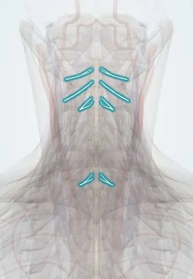
Neck nerve roots !
The cervical nerve roots leave the spinal cord in the neck through side openings; they carry movement and feeling for the neck, arms and hands.
Official name: Anterior root of spinal nerve
Opens in the Durgical app, no sign-up needed.
What it is
The nerve roots of the neck: each leaves the spinal cord, passes through a side opening (foramen) and forms a spinal nerve to the shoulder, arm and hand.
What it does
Carry movement and sensation for the neck, shoulders, arms and hands. C5 to T1 form the brachial plexus.
Key facts
- Roots
- C1–C8 (8 pairs, 7 vertebrae); each exits above its numbered vertebra; C8 exits between C7 and T1
- Moves
- Neck muscles (C1–C4); diaphragm via phrenic nerve (C3–C5); upper limb via brachial plexus (C5–T1)
- Feeling from
- Back of scalp, neck, shoulder and upper limb dermatomes (C1 usually has no dermatome)
Relations
- Course
- Each root pair leaves the cord, crosses the subarachnoid space in a dural sleeve and runs out through the foramen, then along the groove of the transverse process behind the vertebral artery
- In front
- The disc and the uncovertebral joint, whose spurs narrow the foramen from the front; the vertebral artery outside the foramen
- Behind
- The facet joint (superior articular process of the vertebra below)
- Above
- The pedicle of the vertebra above
- Below
- The pedicle of the vertebra below
How to find it
They cannot be felt. In a posterior foraminotomy the root is found where the lamina meets the facet joint, under the medial part of the facet; from the front it lies behind the uncovertebral joint, which marks the lateral limit of an anterior discectomy.
Examination
- Spurling test: neck extended, turned and tilted to the painful side with gentle downward pressure; arm pain in a root pattern supports radiculopathy (specific, not sensitive)
- Shoulder abduction relief sign: resting the hand on top of the head eases the arm pain, suggesting root compression
- Upper limb tension test: stretching the arm's nerves reproduces the symptoms; a negative test makes radiculopathy less likely
- ISNCSCI key muscles: C5 elbow flexors, C6 wrist extensors, C7 elbow extensors, C8 middle-finger flexion at the fingertip, T1 little-finger abduction
On imaging
MRI is the test of choice for radiculopathy: axial T2 shows each root leaving through its foramen and the disc or spur pressing on it; oblique sagittal images show the foramina well. CT shows bony spurs, and CT myelography is used when MRI is not possible. After a brachial plexus traction injury, a pseudomeningocele (fluid pouch at the foramen) on MRI or CT myelography suggests root avulsion.
Common problems
- Cervical radiculopathy: a disc or bone spur pinches a root, causing pain, numbness or weakness down the arm in that root's pattern.
How it is diagnosed
Exam of strength, reflexes and feeling in the arm (each root has its own pattern); MRI; sometimes nerve tests (EMG).
Treatment
- Most improve with time, physical therapy, medicine and sometimes an injection.
- Lasting pain or weakness: surgery to free the root, such as ACDF or a foraminotomy.
Clinical notes
- C8 vs ulnar nerve: both numb the little and ring fingers and weaken the intrinsic hand muscles, but C8 also weakens thumb and index flexion (median) and numbs the medial forearm, which the ulnar nerve does not supply
- Double crush: a root lesion and a distal entrapment can coexist; neck pain and reflex changes point to the root, and nerve conduction studies separate the two
- Anterior cervical discectomy and fusion reaches the disc through the front of the neck, between the carotid sheath laterally and the trachea and oesophagus medially
- Occipital neuralgia: the greater occipital nerve (C2 dorsal ramus) gives shooting pain from the base of the skull over the back of the scalp
Development
The cord stops lengthening early while the column keeps growing. In the neck the mismatch is small, so cervical roots reach their foramina almost horizontally; lower down the roots slope more and more steeply.
Memory aid
"C3, 4, 5 keeps the diaphragm alive": the phrenic nerve, which is why complete injuries at C3 or above usually need a ventilator.
Related conditions
- Cervical radiculopathy
- Guillain-Barré syndrome
- Neurofibromatosis (types 1 and 2)
- Chronic inflammatory demyelinating polyneuropathy (CIDP)
- Cervical Spondylosis
Test yourself
Think of the answer, then tap the question to see it.
Which cervical root does a posterolateral C5–C6 disc herniation compress?
The C6 root: a cervical disc pinches the root numbered for the lower vertebra, since cervical roots exit above their numbered vertebra.
Which cervical nerve root is most often affected in radiculopathy?
C7, then C6; in older people, foraminal narrowing by uncovertebral and facet spurs is a more common cause than soft disc.
What is the Spurling test?
The neck is extended, turned and tilted to the painful side with gentle downward pressure; arm pain in a root pattern supports radiculopathy (specific, not sensitive).
Which cervical roots form the phrenic nerve, and why does it matter in spinal cord injury?
C3, C4 and C5; complete injuries at C3 or above usually need a ventilator.
Educational information, not medical advice. For any symptom, see a health professional.
Its names
- Official Latin (TA2)
- radix anterior nervi spinalis; radix posterior nervi spinalis; nervi cervicales TA2 6145
- Official Spanish (DeCS)
- Raíces nerviosas espinales DeCS D013126
- English (TA2)
- Anterior root of spinal nerve
- Also called
- motor root of spinal nerve · ventral root of spinal nerve · sensory root of spinal nerve · dorsal root of spinal nerve · spinal nerves C1-C8
Related
- Dorsal root ganglia
- Brachial plexus roots
- C5 nerve root
- C6 nerve root
- C7 nerve root
- Longus colli
- Longus capitis
- Rectus capitis anterior and lateralis
Sources: Terminologia Anatomica (TA2); DeCS; Anatomy, Head and Neck: Cervical Nerves; Anatomy, Skin, Dermatomes; 13.2 The Central Nervous System (OpenStax (Rice University), 2022) · adapted, CC BY 4.0.
Spotted a mistake?We are always improving for the community. Tell us what is wrong and our team will review it.Report a mistake
Representative model, for educational purposes only.