Joints and ligaments · Upper limb
SC disc !
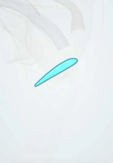
The sternoclavicular disc sits in the joint between the collarbone and the breastbone; it cushions the joint and stops the collarbone sliding inward.
Official name: Sternoclavicular joint disc
Opens in the Durgical app, no sign-up needed.
What it is
A disc of cartilage inside the joint between the collarbone and the breastbone.
What it does
Cushions the joint and stops the collarbone sliding inward.
Key facts
- Attaches
- Upper-back clavicle to the 1st costal cartilage
- Function
- Stops the clavicle sliding medially onto the manubrium; absorbs shock
Relations
- Lateral
- Sternal end of the clavicle
- Medial
- Clavicular notch of the manubrium
- Below
- First costal cartilage at its junction with the manubrium; the costoclavicular ligament and first rib further out
- Above
- SC capsule and the interclavicular ligament
- Behind
- Posterior SC ligament, then sternohyoid and sternothyroid, with the brachiocephalic vein and great vessels beyond
How to find it
The joint lies under the skin beside the jugular notch, lateral to the cord-like sternal head of sternocleidomastoid; with that muscle relaxed, a small hollow between the bulky end of the clavicle and the manubrium marks the joint line.
On imaging
CT is the best study for the SC joint and its relation to the vessels; MRI shows the disc, effusion and the medial physis. On plain X-ray the serendipity view (AP with about 40° of cephalic tilt) compares the two clavicle ends.
Clinical notes
- Septic arthritis of the SC joint is classically seen in people who inject drugs, in diabetes and with central venous lines
- Disc tears or degeneration cause a painful click with shoulder movement; SC osteoarthritis is common with age and often painless
- Inflammatory and sclerosing conditions, such as SAPHO syndrome and condensing osteitis of the clavicle, also involve this joint
Development
Forms from mesenchyme between clavicle and sternum that persists instead of being resorbed when the two cavities open (older texts saw it as the remnant of a lost skeletal element). The secondary centre at the sternal end of the clavicle beside it is the last in the body to unite.
Test yourself
Think of the answer, then tap the question to see it.
How does the sternoclavicular disc divide the SC joint?
Into two separate synovial cavities: clavicle–disc and disc–manubrium.
Thanks to the sternoclavicular disc, which movements does the SC joint allow?
Elevation and depression, protraction and retraction, and rotation of the clavicle, almost like a ball and socket.
In a young adult, what can an apparent sternoclavicular dislocation actually be?
A fracture through the medial clavicular physis, next to the SC disc, the last physis to fuse (early to mid-20s).
Educational information, not medical advice. For any symptom, see a health professional.
Its names
- Official Latin (TA2)
- discus articulationis sternoclavicularis TA2 1751
- Official Spanish (DeCS)
- Disco articular de la articulación esternoclavicular
- English (TA2)
- Sternoclavicular joint disc
Related
Sources: Terminologia Anatomica (TA2); Anatomy, Shoulder and Upper Limb, Sternoclavicular Joint; Anatomy of the Human Body (20th ed.): Surface Anatomy of the Upper Extremity (Lea & Febiger (Gray's Anatomy, 1918), 1918) · adapted, Public domain; Anatomy of the Human Body (20th ed.): Development of the Joints (Lea & Febiger (Gray's Anatomy, 1918), 1918) · adapted, Public domain; Anatomy of the Human Body (20th ed.): The Clavicle (Lea & Febiger (Gray's Anatomy, 1918), 1918) · adapted, Public domain.
Spotted a mistake?We are always improving for the community. Tell us what is wrong and our team will review it.Report a mistake
Representative model, for educational purposes only.