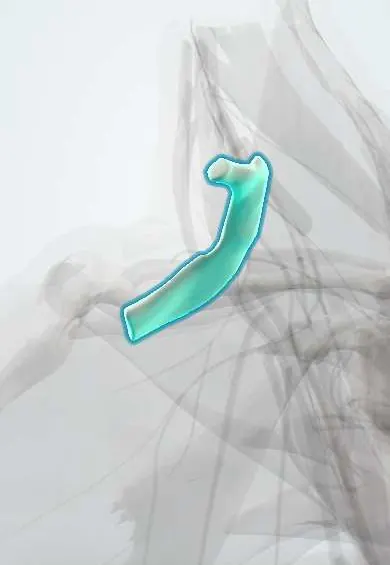
First rib !
The first rib is the shortest, most curved rib, at the base of the neck; the nerves and vessels to the arm cross over it.
Opens in the Durgical app, no sign-up needed.
What it is
The short, curved top rib under the collarbone.
What it does
The brachial plexus and subclavian vessels cross it on their way to the arm.
Key facts
- Joints with
- T1 vertebra (head and tubercle), manubrium (via its cartilage)
- Landmarks
- Scalene tubercle; grooves for the subclavian vein (in front) and artery (behind)
Relations
- Above
- Scalenes: scalenus anterior on the tubercle and scalenus medius behind the groove for the subclavian artery and the lower trunk of the brachial plexus
- In front
- Costoclavicular ligament and subclavius tie it to the clavicle; its cartilage joins the manubrium below the sternoclavicular joint
- Behind
- Head and tubercle against T1; crossing the front of its neck, from medial to lateral: sympathetic trunk, superior intercostal artery and the ventral ramus of T1 rising to join the plexus
- Medial
- Inner border with the suprapleural membrane over the dome of the pleura and the lung apex
How to find it
Hard to feel: it lies behind the clavicle, and only its cartilage is felt just below the sternoclavicular joint at the side of the manubrium. On a chest X-ray it is traced from its posterior end at T1.
On imaging
On a chest or cervical spine X-ray, a cervical rib is told from a T1 rib by its vertebra: C7 transverse processes slope downward, T1 transverse processes slope upward. CT angiography with the arms raised shows vascular thoracic outlet compression.
Clinical notes
- Effort thrombosis of the subclavian vein (Paget–Schroetter syndrome) happens in the costoclavicular space between the first rib and the clavicle, in people who use the arms overhead
- Resection of the first rib is one of the operations for thoracic outlet syndrome
- Stress fractures occur in throwers and rowers; tumours of the lung apex (Pancoast tumour) can invade it with the lower brachial plexus
Anatomical variants
- Cervical rib, usually from C7 and in under 1% of people, on one or both sides; complete or fibrous bands can narrow the thoracic outlet
Development
Ribs start as the costal part of each vertebra's cartilage model, separate from it in the embryo and then ossify, except at the front, where cartilage stays as the costal cartilage. A cervical rib is a costal element of C7 that keeps growing instead of staying small.
Related conditions
Test yourself
Think of the answer, then tap the question to see it.
On the first rib, what does the scalene tubercle separate?
The subclavian vein in front from the subclavian artery and lower trunk of the brachial plexus behind; the scalenus anterior inserts on it.
What does a first rib fracture signal?
High-energy trauma: injuries of the subclavian vessels and brachial plexus are looked for.
Where does effort thrombosis of the subclavian vein (Paget–Schroetter syndrome) occur?
In the costoclavicular space between the first rib and the clavicle, in people who use the arms overhead.
On X-ray, how is a cervical rib told apart from a first rib?
By its vertebra: C7 transverse processes slope downward, T1 transverse processes slope upward.
Educational information, not medical advice. For any symptom, see a health professional.
Its names
- Official Latin (TA2)
- costa prima TA2 1107
- Official Spanish (DeCS)
- Primera costilla
- English (TA2)
- First rib
- Also called
- rib 1
Related
Sources: Terminologia Anatomica (TA2); Anatomy, Thorax, Wall; Anatomy, Thorax, Cervical Rib; 7.5 Embryonic Development of the Axial Skeleton (OpenStax (Rice University), 2022) · adapted, CC BY 4.0.
Spotted a mistake?We are always improving for the community. Tell us what is wrong and our team will review it.Report a mistake
Representative model, for educational purposes only.