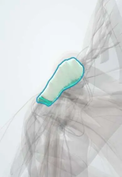
Manubrium !
The manubrium is the top part of the breastbone (sternum); the collarbones join it, making it the inner anchor of the shoulder girdle.
Official name: Manubrium of the sternum
Opens in the Durgical app, no sign-up needed.
What it is
The top part of the breastbone, where the collarbone meets it.
What it does
Forms the inner end of the shoulder girdle (the sternoclavicular joint).
Key facts
- Joints with
- Clavicles (SC joints), 1st costal cartilages, part of the 2nd, body of the sternum
- Landmarks
- Jugular notch, clavicular notches, sternal angle (T4–T5 level, 2nd rib)
Relations
- Above
- Jugular notch, with the trachea above and behind it
- Lateral
- Clavicular notches with the SC capsule, SC ligaments and SC disc; first costal cartilage just below
- Behind
- Sternohyoid and sternothyroid; behind them the left brachiocephalic vein, then the aortic arch with its branches and the thymic remnant
- In front
- Sternal head of the sternocleidomastoid and the origin of the pectoralis major
- Below
- Body of the sternum at the manubriosternal joint, with half of the 2nd costal cartilage
How to find it
The jugular notch is felt at the base of the neck between the clavicular heads; sliding down a few centimetres, the transverse ridge of the sternal angle is felt, with the 2nd costal cartilage beside it, the starting point for counting ribs.
On imaging
Sternoclavicular joints are hard to see on plain films (a view with the beam tilted about 40° toward the head helps); CT is the standard for sternoclavicular dislocation and manubrial fractures.
Clinical notes
- Septic arthritis of the sternoclavicular joint is classically seen in people who inject drugs, people with diabetes and people with central lines
- A median sternotomy splits the manubrium and body to reach the heart and mediastinum
Development
Ossifies from one or more centres in late fetal life; the manubriosternal joint is usually a fibrocartilaginous joint and may stay unfused into old age.
Related conditions
Test yourself
Think of the answer, then tap the question to see it.
What lies at the level of the sternal angle (manubriosternal joint)?
The 2nd costal cartilage and the T4–T5 plane, where the aortic arch begins and ends and the trachea divides.
Which is the only bony joint linking the upper limb to the trunk?
The sternoclavicular joint between the clavicle and the manubrium, a saddle-type synovial joint divided by the SC disc.
Which vein crosses behind the manubrium?
The left brachiocephalic vein, behind sternohyoid and sternothyroid, with the aortic arch and its branches beyond.
Which imaging study is the standard for sternoclavicular dislocation and manubrial fractures?
CT, because these joints are hard to see on plain films.
Educational information, not medical advice. For any symptom, see a health professional.
Its names
- Official Latin (TA2)
- manubrium sterni TA2 1130
- Official Spanish (DeCS)
- Manubrio del esternón
- English (TA2)
- Manubrium of the sternum
Related
Sources: Terminologia Anatomica (TA2); Anatomy, Thorax, Sternum.
Spotted a mistake?We are always improving for the community. Tell us what is wrong and our team will review it.Report a mistake
Representative model, for educational purposes only.