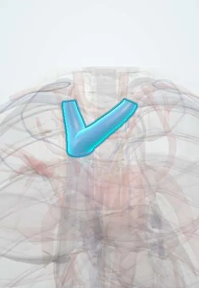
Brachiocephalic veins !
The brachiocephalic veins form behind each collarbone and drain the head, neck and arms; they join to form the superior vena cava.
Official name: Brachiocephalic vein
Opens in the Durgical app, no sign-up needed.
What it is
Two veins behind the top of the sternum, formed where the jugular and subclavian veins join; they merge into the superior vena cava.
What it does
Drain the head, neck and arms.
Key facts
- Drains
- Internal jugular and subclavian veins; vertebral, internal thoracic, inferior thyroid veins; lymph ducts at venous angles
- Into
- Superior vena cava, uniting behind right 1st costal cartilage; left vein longer, crosses behind manubrium
Relations
- Course
- Right: descends almost straight down from behind the right sternoclavicular joint. Left: runs down and to the right behind the upper half of the manubrium. They meet behind the right 1st costal cartilage
- In front
- Manubrium and sternoclavicular joints, the origins of the sternohyoid and sternothyroid muscles, and the thymus or its fatty remnant
- Behind
- Left vein: the brachiocephalic trunk, left common carotid and left subclavian arteries and the trachea. Right vein: the right vagus nerve, with the right phrenic nerve and pleura along its side
- Below
- Left vein: the upper border of the aortic arch
How to find it
Not palpable. Each vein starts behind the sternoclavicular joint of its own side. The right one drops almost straight down beside the sternal edge; the left one runs obliquely behind the upper manubrium to meet it behind the right 1st costal cartilage, where the superior vena cava begins.
On imaging
On contrast CT the left vein crosses obliquely behind the manubrium, in front of the arch branches, to join the short right vein as the superior vena cava. CT venography shows clot or narrowing around catheters and pacemaker leads as a filling defect with collaterals. On a chest radiograph a left central line crosses the midline along this vein.
Common problems
- Superior vena cava syndrome: blockage by a tumour or clot, causing swelling of the face and arms.
Clinical notes
- Blockage of one vein (clot around a catheter or pacemaker lead, or tumour) swells the face, neck and arm on that side only, unlike superior vena cava obstruction
- Lying just behind the manubrium, the left vein can bleed heavily when the sternum is reopened or the upper mediastinum is dissected
- The thoracic duct enters at the left venous angle and the right lymphatic duct at the right one; dissection low in the left neck can cause a chyle leak
- Valveless and held open by the fascia of the root of the neck, they do not collapse when cut: an open wound or an open central line here can suck in air (venous air embolism)
Anatomical variants
- Left brachiocephalic vein passing below the aortic arch instead of in front of its branches (rare)
- With a persistent left superior vena cava the left brachiocephalic vein is often small or absent
Development
The left vein forms from an oblique anastomosis between the two anterior cardinal veins; the left anterior cardinal vein below it normally regresses, and if it persists it becomes a left superior vena cava.
Test yourself
Think of the answer, then tap the question to see it.
How do the right and left brachiocephalic veins differ?
The left is much longer and runs obliquely behind the upper manubrium; the right is short and almost vertical. They join behind the right 1st costal cartilage.
Which arteries does the left brachiocephalic vein cross in front of?
The three branches of the aortic arch: brachiocephalic trunk, left common carotid and left subclavian arteries.
How does blockage of one brachiocephalic vein differ clinically from superior vena cava obstruction?
It swells the face, neck and arm on that side only.
Which lymphatic ducts enter at the venous angles of the brachiocephalic veins?
The thoracic duct at the left venous angle and the right lymphatic duct at the right one.
Educational information, not medical advice. For any symptom, see a health professional.
Its names
- Official Latin (TA2)
- vena brachiocephalica TA2 4772
- Official Spanish (DeCS)
- Venas braquiocefálicas DeCS D016121
- English (TA2)
- Brachiocephalic vein
- Also called
- innominate vein · vena anonyma
Related
Sources: Terminologia Anatomica (TA2); DeCS; Anatomy, Thorax, Brachiocephalic (Innominate) Veins; Anatomy of the Human Body (20th ed.): Surface Markings of the Thorax (Lea & Febiger (Gray's Anatomy, 1918), 1918) · adapted, Public domain; Tratado de anatomía humana (9.ª ed.), tomo II, Libro IV Angiología, sección 4: Venas (sistemas de las venas cavas superior e inferior, vena porta, venas del raquis).
Spotted a mistake?We are always improving for the community. Tell us what is wrong and our team will review it.Report a mistake
Representative model, for educational purposes only.