Joints and ligaments · Upper limb
SC ligaments !
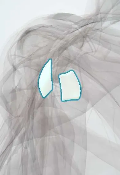
The sternoclavicular ligaments hold the collarbone to the breastbone, the only bony link between the arm and the trunk.
Official name: Sternoclavicular ligaments
Opens in the Durgical app, no sign-up needed.
What it is
Bands in front of and behind the joint between the collarbone and the breastbone.
What it does
Hold the only bony link between the arm and the trunk.
Key facts
- Attaches
- Sternal end of the clavicle to the manubrium (anterior and posterior)
- Function
- Stabilise the only bony joint between arm and trunk; posterior is the strongest
- Tight in
- Translation of the medial clavicle
Relations
- In front
- Origins of the sternocleidomastoid and the clavicular head of the pectoralis major
- Behind
- Sternohyoid and sternothyroid, then the brachiocephalic vein, common carotid artery, trachea and oesophagus (on the right, the brachiocephalic trunk)
- Above
- Interclavicular ligament across the jugular notch
- Below
- Costoclavicular ligament and the first rib
- Deep
- SC disc and the two joint cavities
How to find it
The sternal end of the clavicle stands out beside the jugular notch; it rises slightly when the shoulders shrug and glides when they are pulled back. An anterior dislocation makes it prominent; a posterior one leaves a hollow, which swelling can hide.
On imaging
Plain AP films are hard to read here; the serendipity view (about 40° of cephalic tilt) helps, but CT, with contrast when vessels may be injured, is the study of choice to show the direction of displacement and any compression of the mediastinum.
Clinical notes
- Posterior dislocation can cause shortness of breath, difficulty swallowing, voice change or arm swelling from venous compression; reduction is done with vascular or thoracic surgical support available
- Chronic instability is treated with a tendon graft in a figure-of-eight between clavicle and manubrium; any resection of the medial clavicle preserves the costoclavicular ligament
- Anterior dislocations are usually managed without surgery; a lasting bump over the joint is common but rarely limits function
Test yourself
Think of the answer, then tap the question to see it.
Which sternoclavicular ligament resists both forward and backward translation of the medial clavicle?
The posterior SC ligament, the thickened posterior capsule.
Which sternoclavicular dislocation is more common and which is an emergency?
Anterior is far more common and usually benign; posterior is rarer but an emergency, pressing on great vessels, trachea and oesophagus.
Which imaging study is the choice for a suspected SC ligament injury?
CT, with contrast when vessels may be injured, to show the direction of displacement.
Which symptoms suggest compression after a posterior sternoclavicular dislocation?
Shortness of breath, difficulty swallowing, voice change or arm swelling from venous compression.
Educational information, not medical advice. For any symptom, see a health professional.
Its names
- Official Latin (TA2)
- ligamentum sternoclaviculare anterius TA2 1754
- Official Spanish (DeCS)
- Ligamentos esternoclaviculares anterior y posterior
- English (TA2)
- Sternoclavicular ligaments
Related
Sources: Terminologia Anatomica (TA2); Anatomy, Shoulder and Upper Limb, Sternoclavicular Joint.
Spotted a mistake?We are always improving for the community. Tell us what is wrong and our team will review it.Report a mistake
Representative model, for educational purposes only.