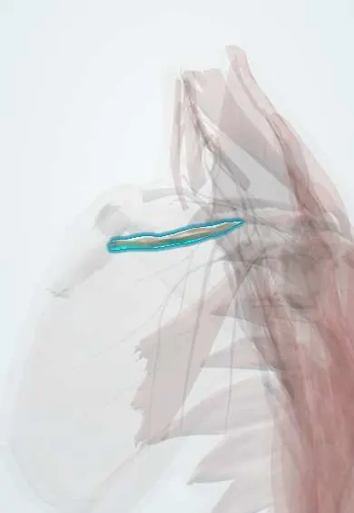
Subclavius !
The subclavius is a small muscle under the collarbone; it steadies the collarbone against the breastbone and cushions the vessels below.
Opens in the Durgical app, no sign-up needed.
What it is
A small muscle under the collarbone.
What it does
Steadies the collarbone against the breastbone.
Key facts
- Origin
- Junction of the 1st rib and its cartilage
- Insertion
- Inferior surface of the middle third of the clavicle
- Action
- Anchors and depresses the clavicle
- Nerve
- Nerve to subclavius (C5–C6)
- Blood supply
- Thoracoacromial artery (clavicular branch)
Relations
- Above
- The groove on the underside of the middle third of the clavicle
- Below
- The subclavian vein, with the subclavian artery and the brachial plexus behind it, all crossing the first rib
- In front
- The clavipectoral fascia and the clavicular head of the pectoralis major
- Medial
- The costoclavicular ligament, near its origin at the first rib and its cartilage
How to find it
Not palpable: it hides in the groove under the clavicle. In dissection it appears on dividing the clavipectoral fascia below the clavicle; on ultrasound it is the thin band between the clavicle and the subclavian vein.
Examination
- Costoclavicular (military brace) test: the shoulders are drawn back and down while the radial pulse is felt; loss of the pulse or reproduced symptoms suggests compression between the clavicle with the subclavius and the first rib, though many healthy people also lose the pulse
On imaging
A small muscle under the clavicle, seen on CT or MRI between the bone and the subclavian vein. In venous thoracic outlet syndrome, studies with the arm raised (Doppler ultrasound, CT or MR venography) show the vein narrowed in the costoclavicular space; Doppler ultrasound is the first test for subclavian vein thrombosis.
Clinical notes
- Drilling and plating the clavicle put the subclavian vessels at risk; the subclavius is the only soft tissue between the bone and the vein
- Surgery for venous thoracic outlet syndrome resects the first rib and divides the subclavius tendon and costoclavicular ligament to free the vein
Anatomical variants
- An accessory phrenic nerve often arises from the nerve to subclavius and joins the phrenic nerve in the chest (frequency varies between studies)
Related conditions
Test yourself
Think of the answer, then tap the question to see it.
What does the subclavius do in a midshaft clavicle fracture?
Lying between the clavicle and the subclavian vein, it partly cushions the subclavian vessels and the brachial plexus from the bone ends.
How can the subclavius cause venous thoracic outlet syndrome?
Its tendon, with the costoclavicular ligament and the first rib, can squeeze the subclavian vein in the costoclavicular space (effort thrombosis).
What are the attachments and nerve supply of the subclavius?
From the junction of the 1st rib and its cartilage to the underside of the middle third of the clavicle; nerve to subclavius (C5–C6).
Educational information, not medical advice. For any symptom, see a health professional.
Its names
- Official Latin (TA2)
- musculus subclavius TA2 2306
- Official Spanish (DeCS)
- Músculo subclavio
- English (TA2)
- Subclavius
Related
Sources: Terminologia Anatomica (TA2); Anatomy, Shoulder and Upper Limb, Pectoral Muscles.
Spotted a mistake?We are always improving for the community. Tell us what is wrong and our team will review it.Report a mistake
Representative model, for educational purposes only.