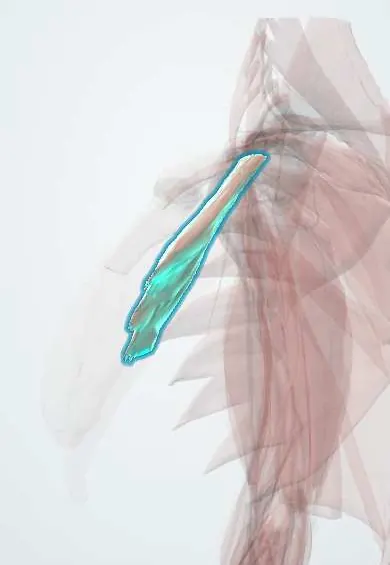
Pectoralis minor !
The pectoralis minor is a thin chest muscle under the pectoralis major; it pulls the shoulder blade forward, and the arm's nerves and vessels pass under it.
Opens in the Durgical app, no sign-up needed.
What it is
A thin triangular muscle under the pectoralis major, from the ribs to the coracoid.
What it does
Pulls the shoulder blade forward and down; the plexus and vessels pass under it.
Key facts
- Origin
- Ribs 3–5 near their costal cartilages (varies: 2–5)
- Insertion
- Coracoid process of scapula (medial border and upper surface)
- Action
- Stabilizes scapula, pulling it forward and down against chest wall; raises ribs in forced inspiration if scapula fixed
- Nerve
- Medial pectoral nerve (C8–T1) (some texts: also lateral pectoral nerve)
- Blood supply
- Pectoral branch of thoracoacromial artery; lateral thoracic artery
Relations
- Superficial
- Pectoralis major and the lateral pectoral nerve
- Deep
- Second part of the axillary artery, the cords of the brachial plexus and the axillary vein, then the ribs and serratus anterior
- Above
- The clavipectoral fascia, stretched between its upper border, the subclavius and the clavicle; the cephalic vein, thoracoacromial artery and lateral pectoral nerve pass through it
- Below
- The lateral thoracic artery runs along its lower border; the medial pectoral nerve pierces the muscle on its way to pectoralis major
How to find it
Not palpable as a separate muscle: it lies under the pectoralis major, fanning from the coracoid process down to ribs 3–5. It is exposed by reflecting the pectoralis major in dissection, and on ultrasound it is the thin layer between the pectoralis major and the axillary vessels below the coracoid.
Examination
- Hyperabduction (Wright) test: loss of the radial pulse or reproduced tingling with the arm abducted overhead suggests compression under the pectoralis minor or in the costoclavicular space; many healthy people also lose the pulse
- Pectoralis minor length: lying supine, a shoulder that rests higher off the table than the other suggests a short muscle tipping the scapula forward
On imaging
On axial CT or MRI it runs from the 3rd to 5th ribs up to the coracoid, in front of the axillary vessels. On breast cancer staging scans it is the landmark for axillary node levels: lateral (I), behind (II) and medial (III). Ultrasound shows it deep to the pectoralis major and guides the pectoral nerve blocks.
Clinical notes
- Pectoralis minor syndrome: the brachial plexus and axillary vessels are compressed beneath it during overhead positions; release of the tendon is one treatment
- A tight muscle tips the scapula forward and narrows the subacromial space, a common finding in scapular dyskinesis
- Pectoral nerve (PECS) blocks place local anaesthetic between the pectoralis major and minor, and between the minor and serratus anterior, for breast and chest wall surgery
Anatomical variants
- Part of its tendon may pass over the coracoid process to join the coracohumeral ligament or the shoulder capsule instead of stopping on the coracoid
- It may be small or absent together with the sternocostal head of the pectoralis major in Poland syndrome
Development
With the pectoralis major it arises next to the arm bud while the bud still lies at lower cervical levels and then spreads down over the chest, which is why branches of the brachial plexus (medial and lateral pectoral nerves) supply it.
Memory aid
Axillary artery: part 1 has 1 branch, part 2 has 2, part 3 has 3. 'Screw The Lawyer, Save A Patient': superior thoracic; thoracoacromial, lateral thoracic; subscapular, anterior and posterior circumflex humeral.
Related conditions
Test yourself
Think of the answer, then tap the question to see it.
How does the pectoralis minor divide the axillary artery?
It crosses in front of it, making three parts: medial to the muscle, behind it and lateral to it.
How are axillary lymph node levels defined by the pectoralis minor?
Level I lateral to (below) the muscle, level II behind it, level III medial to (above) it.
What is pectoralis minor syndrome?
Compression of the brachial plexus and axillary vessels beneath the muscle in overhead positions; tendon release is one treatment.
Where does the pectoralis minor attach, and what nerve supplies it?
From ribs 3–5 to the coracoid process; medial pectoral nerve (C8–T1).
Educational information, not medical advice. For any symptom, see a health professional.
Its names
- Official Latin (TA2)
- musculus pectoralis minor TA2 2305
- Official Spanish (DeCS)
- Músculo pectoral menor
- English (TA2)
- Pectoralis minor
Related
- Axillary artery
- Anterior axillary nodes
- Interpectoral nodes
- Pectoral nerves
- Thoracoacromial artery
- Lateral thoracic artery
- Scapula
Sources: Terminologia Anatomica (TA2); Anatomy, Shoulder and Upper Limb, Pectoral Muscles; Anatomy of the Human Body (20th ed.): Development of the Muscles (Lea & Febiger (Gray's Anatomy, 1918), 1918) · adapted, Public domain.
Spotted a mistake?We are always improving for the community. Tell us what is wrong and our team will review it.Report a mistake
Representative model, for educational purposes only.