Cardiovascular system · Upper limb
Subclavian artery !
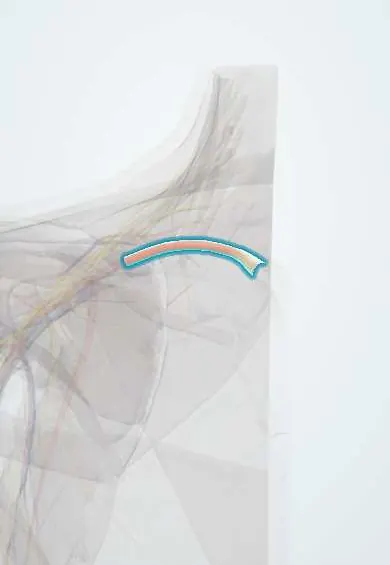
The subclavian artery arches over the first rib behind the collarbone; it brings blood to the arm and becomes the axillary artery past the rib.
Opens in the Durgical app, no sign-up needed.
What it is
The large artery that arches over the first rib behind the collarbone.
What it does
Brings blood to the arm; past the first rib it becomes the axillary artery.
Key facts
- Origin
- Right: brachiocephalic trunk; left: aortic arch; 3 parts relative to anterior scalene
- Branches
- 1st part: vertebral, internal thoracic, thyrocervical trunk (left: also costocervical trunk); 2nd part: costocervical trunk (right); 3rd part: dorsal scapular (often)
- Supplies
- Upper limb, brain (vertebral), neck, chest wall; becomes axillary artery at lateral border of 1st rib
Relations
- Course
- Right: from the brachiocephalic trunk behind the right sternoclavicular joint; left: from the aortic arch, rising through the superior mediastinum. Each arches over the cervical pleura, passes between the anterior and middle scalenes and crosses the first rib behind the clavicle
- In front
- The anterior scalene with the phrenic nerve on it; in front of that the subclavian vein, the subclavius and the clavicle; on the right the vagus nerve crosses the first part and its recurrent laryngeal branch hooks under it
- Behind
- Behind the first part: the cervical pleura, the ansa subclavia and the cervicothoracic (stellate) ganglion, and on the right the recurrent laryngeal nerve; then the brachial plexus trunks: upper and middle above and behind it, the lower trunk directly behind its third part on the first rib
- Below
- Dome of the pleura and apex of the lung under the first part; the shallow groove on the upper surface of the first rib under the third part
- Medial
- Left artery in the superior mediastinum: trachea, oesophagus and the left recurrent laryngeal nerve medially, left pleura and lung laterally; at the root of the neck the thoracic duct arches over its first part to reach the left venous angle
How to find it
The third part is felt in the supraclavicular fossa, just above the middle of the clavicle, by pressing downward and backward against the first rib. A bruit heard there can point to a narrowing. On ultrasound it lies above the clavicle, with the vein in front of the scalene muscle.
Examination
- Adson test: with the radial pulse monitored, the seated person breathes in deeply, extends the neck and turns the chin toward the tested side; a fading pulse or reproduced symptoms suggests compression in the scalene gap, though many healthy people also test positive
- Wright (hyperabduction) test: the arm is raised overhead in abduction and external rotation while the radial pulse is felt; loss of the pulse with symptoms points to compression under pectoralis minor or in the costoclavicular space, with frequent false positives
On imaging
Duplex ultrasound shows flow; in subclavian steal the vertebral artery on the same side shows reversed flow. CT or MR angiography maps stenosis, aneurysm or compression in thoracic outlet syndrome, with images taken with the arm down and raised. A chest radiograph may reveal a cervical rib. A blood pressure difference between the arms is a common reason to image it.
Clinical notes
- Arterial thoracic outlet syndrome: a cervical rib or a tight scalene gap compresses the third part, causing narrowing, a dilatation beyond it and clots that travel to the fingers
- A clear difference in blood pressure between the two arms suggests a narrowed subclavian artery on the lower side
- Its branch, the internal thoracic artery, is the classic coronary bypass conduit; a narrowing of the subclavian before that branch can rob the graft (coronary-subclavian steal)
- During subclavian vein lines the artery lies just behind and above the vein; an arterial puncture there is hard to compress because the clavicle is in the way
Anatomical variants
- Aberrant right subclavian artery (arteria lusoria): the most common aortic arch anomaly; it arises as the last branch of the arch and usually passes behind the esophagus, mostly silent but a cause of dysphagia lusoria
- The dorsal scapular artery may come from the subclavian artery or from the transverse cervical artery; the left vertebral artery sometimes arises directly from the aortic arch
Development
The right subclavian comes from the right fourth pharyngeal arch artery plus the seventh intersegmental artery; the left from the left seventh intersegmental artery alone. Loss of the right fourth arch segment leaves the aberrant right subclavian.
Memory aid
Branches in order: "VIT C, D" (Vertebral, Internal thoracic, Thyrocervical, Costocervical, Dorsal scapular).
Related conditions
Test yourself
Think of the answer, then tap the question to see it.
Which muscle divides the subclavian artery into three parts?
The anterior scalene: medial to, behind and lateral to the muscle; the subclavian vein passes in front of it.
Where can the subclavian artery be compressed to slow bleeding from the arm?
Its third part, pressed down onto the first rib in the supraclavicular fossa.
What is subclavian steal?
A narrowing of the subclavian artery before the vertebral artery's origin lets the arm draw blood backwards down the vertebral artery, worse with arm exercise.
What is an aberrant right subclavian artery (arteria lusoria)?
The most common aortic arch anomaly: it arises as the last branch of the arch and usually passes behind the esophagus; it can cause dysphagia lusoria.
What does a clear blood pressure difference between the two arms suggest?
A narrowed subclavian artery on the side with the lower pressure.
Educational information, not medical advice. For any symptom, see a health professional.
Its names
- Official Latin (TA2)
- arteria subclavia TA2 4537
- Official Spanish (DeCS)
- Arteria subclavia
- English (TA2)
- Subclavian artery
Related
Sources: Terminologia Anatomica (TA2); Anatomy, Thorax, Subclavian Arteries; Tratado de anatomía humana (9.ª ed.), tomo II, Libro IV Angiología, arterias subclavia, axilar, humeral, radial y cubital, arcos palmares y vías anastomóticas del miembro superior.
Spotted a mistake?We are always improving for the community. Tell us what is wrong and our team will review it.Report a mistake
Representative model, for educational purposes only.