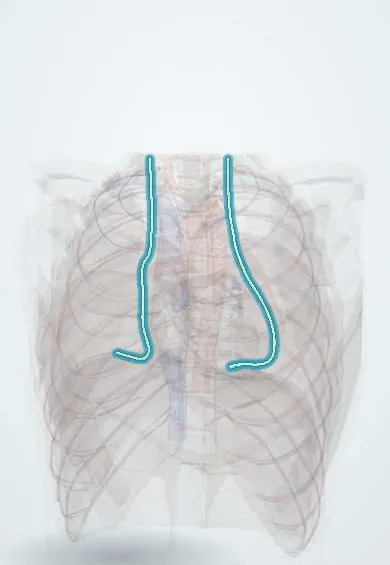
Phrenic nerves !
The phrenic nerves run from the neck down each side of the heart to the diaphragm; they are the only nerves that make it contract for breathing.
Official name: Phrenic nerve
Opens in the Durgical app, no sign-up needed.
What it is
Two nerves from the neck (C3, C4, C5) that run down over the heart's covering to the diaphragm.
What it does
The only nerves that make the diaphragm contract: every breath starts with them.
Key facts
- Roots
- C3–C5 (mainly C4); descend on anterior scalene, then in front of lung root on pericardium
- Moves
- Diaphragm (only motor supply)
- Feeling from
- Mediastinal and diaphragmatic pleura, pericardium, central diaphragm peritoneum; pain referred to shoulder (C4)
Relations
- Course
- In the neck it descends on the front of the anterior scalene under the prevertebral fascia, enters the chest between the subclavian artery and vein, crosses the internal thoracic artery, then runs down on the pericardium in front of the lung root to the diaphragm
- In front
- In the neck: the sternocleidomastoid, the internal jugular vein and the transverse cervical and suprascapular arteries
- Behind
- Anterior scalene muscle in the neck; the lung root in the chest
- Medial
- Right: the right brachiocephalic vein, superior vena cava and right atrium. Left: the left common carotid and subclavian arteries, the aortic arch and the left ventricle
- Lateral
- Mediastinal pleura and lung
How to find it
In the neck it is traced on the front of the anterior scalene, deep to the posterior border of the sternocleidomastoid at about cricoid level, and can be seen there on ultrasound; at surgery in the chest it runs down on the pericardium in front of the lung root.
Examination
- Sniff test (fluoroscopy or ultrasound): a paralysed hemidiaphragm moves up instead of down on a quick sniff
- Diaphragm ultrasound: little or no thickening of the diaphragm muscle during inspiration on the affected side
- Phrenic nerve conduction study from the neck: absent or delayed diaphragm response points to nerve injury
On imaging
Not seen on routine imaging; it is judged by diaphragm movement. A raised hemidiaphragm on the chest radiograph suggests palsy. Ultrasound or the fluoroscopic sniff test confirms it: on a quick sniff the paralysed side fails to descend or moves up. CT then follows the nerve's course through the neck and mediastinum for a tumour or nodes.
Common problems
- Injury paralyses half of the diaphragm: breathlessness, especially lying flat.
- Irritation causes hiccups or pain felt in the shoulder tip.
How it is diagnosed
Chest X-ray (raised diaphragm), ultrasound or fluoroscopy 'sniff test'.
Treatment
- Often recovers; surgical plication of the diaphragm in some cases.
Clinical notes
- An interscalene brachial plexus block almost always reaches the phrenic nerve, briefly paralysing the hemidiaphragm on that side
- Injured in heart surgery (cold injury, internal thoracic artery harvest), in lung and mediastinal surgery and in thymectomy; invaded by lung cancer or pressed by mediastinal masses
- Spinal cord injury at or above C3 abolishes diaphragm breathing; injury below C5 spares it
- Phrenic nerve pacing can support breathing in selected high spinal cord injuries
Anatomical variants
- Accessory phrenic nerve, often from the nerve to subclavius (C5), joining it low in the neck or upper chest; it can escape a block or keep part of the diaphragm working after injury
Development
The diaphragm muscle forms in the neck region, in the septum transversum, and descends to the upper abdomen as the embryo grows, pulling its C3–C5 nerve along; this explains the long course and why diaphragmatic irritation is felt at the shoulder.
Memory aid
C3, 4 and 5 keep the diaphragm alive.
Related conditions
Test yourself
Think of the answer, then tap the question to see it.
What are the roots of the phrenic nerve and what is its motor supply?
C3–C5, mainly C4; it is the only motor supply to the diaphragm.
Do the phrenic nerves pass in front of or behind the lung roots?
In front, between the fibrous pericardium and mediastinal pleura with the pericardiacophrenic vessels; the vagus nerves pass behind.
What is the sniff test for phrenic nerve palsy?
On fluoroscopy or ultrasound, a paralysed hemidiaphragm moves up instead of down during a quick sniff.
Why is pain from diaphragmatic irritation felt at the shoulder tip?
The phrenic nerve carries sensory fibres from the central diaphragm and pleura, and their pain is referred to the shoulder through C4.
Which regional anaesthetic block almost always reaches the phrenic nerve?
The interscalene brachial plexus block, briefly paralysing the hemidiaphragm on that side.
Educational information, not medical advice. For any symptom, see a health professional.
Its names
- Official Latin (TA2)
- nervus phrenicus TA2 6380
- Official Spanish (DeCS)
- Nervio frénico DeCS D010791
- English (TA2)
- Phrenic nerve
- Also called
- bell’s internal respiratory nerve
Related
Sources: Terminologia Anatomica (TA2); DeCS; Anatomy, Thorax, Phrenic Nerves; Anatomy of the Human Body (20th ed.): Development of the Body Cavities (Lea & Febiger (Gray's Anatomy, 1918), 1918) · adapted, Public domain.
Spotted a mistake?We are always improving for the community. Tell us what is wrong and our team will review it.Report a mistake
Representative model, for educational purposes only.