Cardiovascular system · Upper limb
Subclavian vein !
The subclavian vein runs under the collarbone in front of the first rib; it drains the arm toward the heart and is a common route for central lines.
Opens in the Durgical app, no sign-up needed.
What it is
The large vein under the collarbone, in front of the artery.
What it does
Drains the arm toward the heart; a common route for central lines.
Key facts
- Drains
- Upper limb (continues axillary vein at lateral border of 1st rib); external jugular vein; runs in front of anterior scalene
- Into
- Brachiocephalic vein, joining internal jugular vein behind sternoclavicular joint
Relations
- Course
- From the outer border of the first rib it arches over the rib in front of the anterior scalene and behind the clavicle, ending behind the sternoclavicular joint
- In front
- Clavicle and subclavius
- Behind
- Anterior scalene with the phrenic nerve, and behind the muscle the subclavian artery
- Below
- First rib, and medially the dome of the pleura and the apex of the lung
- Medial
- The costoclavicular ligament, which closes the costoclavicular space on the inner side
How to find it
Not palpable. For central lines it is reached below the clavicle near the junction of its medial and middle thirds, or above the clavicle beside the clavicular head of sternocleidomastoid; ultrasound guidance is now usual.
On imaging
On ultrasound it is a thin-walled vein that changes calibre with breathing, beside the pulsating artery; venography or CT with the arm raised can show compression in the costoclavicular space.
Common problems
- A common route for central venous catheters and pacemaker leads.
- Effort thrombosis (Paget-Schroetter): a clot from compression between the collarbone and first rib in heavy overhead users.
- Clots related to catheters.
How it is diagnosed
A swollen, heavy, bluish arm; ultrasound or venography confirms a clot.
Treatment
- Blood thinners; sometimes clot-dissolving treatment.
- Surgery to relieve the outlet compression in effort thrombosis.
Clinical notes
- Subclavian central lines: the pleura lies just below and behind, so pneumothorax is a recognised risk, and an arterial puncture is hard to compress under the clavicle
- Pinch-off: a catheter entering far medially between the clavicle and the first rib can be compressed and eventually break
- Effort thrombosis (Paget-Schroetter) follows repeated compression in the costoclavicular space; this anatomy explains why some treatments remove part of the first rib
- Left-sided lines pass close to the thoracic duct at the venous angle
Anatomical variants
- The vein may lie higher than usual or pass behind the anterior scalene together with the artery
Related conditions
Test yourself
Think of the answer, then tap the question to see it.
How does the subclavian vein relate to the anterior scalene and the subclavian artery?
The vein lies in front of the anterior scalene, the artery behind it; the phrenic nerve runs on the muscle between them.
What is effort thrombosis (Paget-Schroetter) of the subclavian vein?
A clot from repeated compression of the vein in the costoclavicular space, between the clavicle and the first rib, in heavy overhead users.
What is catheter pinch-off in subclavian lines?
A catheter entering far medially between the clavicle and first rib can be compressed and eventually break.
Which lymphatic ducts join at the venous angle formed by the subclavian vein?
The thoracic duct on the left and the right lymphatic duct on the right.
Educational information, not medical advice. For any symptom, see a health professional.
Its names
- Official Latin (TA2)
- vena subclavia TA2 4953
- Official Spanish (DeCS)
- Vena subclavia
- English (TA2)
- Subclavian vein
Related
Sources: Terminologia Anatomica (TA2); Anatomy, Thorax, Subclavian Veins.
Spotted a mistake?We are always improving for the community. Tell us what is wrong and our team will review it.Report a mistake
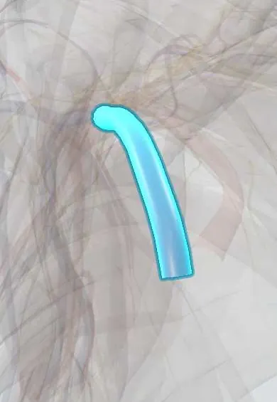
Representative model, for educational purposes only.