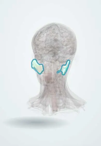
Digestive system · Head and neck
Parotid gland !
The parotid is the largest salivary gland, in front of the ear over the jaw angle; it makes saliva, and the facial nerve branches run through it.
Opens in the Durgical app, no sign-up needed.
What it is
The largest salivary gland, in front of the ear over the jaw angle.
What it does
Makes saliva; the facial nerve branches run through it.
Key facts
- Where
- In front of and below the ear, over the masseter; duct opens opposite the upper 2nd molar
- Blood supply
- External carotid branches (transverse facial)
- Nerve
- Parasympathetic: IX via the otic ganglion and auriculotemporal nerve; facial nerve passes through (not supplying it)
- Function
- Serous saliva
Relations
- Superficial
- Skin, the tough parotid fascia (from the investing cervical fascia) and superficial lymph nodes; the great auricular nerve reaches its lower part
- Deep
- Styloid process and its muscles, the posterior belly of the digastric, the internal jugular vein and the internal carotid artery; the deep lobe reaches the parapharyngeal space
- In front
- Masseter and the ramus of the mandible, which the gland wraps; the duct runs forward over the masseter
- Behind
- Mastoid process and the sternocleidomastoid; the external acoustic meatus above
How to find it
Felt as soft tissue in front of the tragus and over the angle of the jaw. The parotid (Stensen) duct can be rolled under a finger across the clenched masseter, on the middle third of a line from the tragus to midway between the nasal ala and the upper lip.
Examination
- Minor starch–iodine test: iodine painted on the cheek and dusted with starch turns dark blue where sweat appears while eating, confirming Frey syndrome
- Massaging the gland toward the duct opening, on the cheek opposite the upper second molar: pus at the opening suggests bacterial parotitis
On imaging
Ultrasound comes first: the superficial lobe looks evenly bright because of its fat. It shows stones, abscess and solid masses, such as the well-defined, lobulated pleomorphic adenoma, and guides needle biopsy. MRI is chosen for large or deep-lobe tumours, to place them against the facial nerve plane (landmark: retromandibular vein) and to look for perineural spread. CT suits abscess and stones.
Common problems
- Mumps and bacterial infection (parotitis): painful swelling in front of the ear.
- Tumours, most benign (pleomorphic adenoma).
How it is diagnosed
Exam, ultrasound or MRI and a needle biopsy.
Treatment
- Fluids, pain relief, antibiotics for bacterial infection.
- Parotidectomy with careful protection of the facial nerve.
Clinical notes
- In parotidectomy the facial nerve trunk is found with fixed landmarks: the tragal pointer, the posterior belly of the digastric and the tympanomastoid suture; temporary facial weakness afterwards is common
- Frey syndrome (gustatory sweating): after parotid surgery, cut parasympathetic fibres in the auriculotemporal nerve regrow into skin sweat glands, so the cheek sweats and flushes when eating
- Facial weakness with a parotid lump points to a malignant tumour until proven otherwise
- Mumps typically swells both parotids and lifts the ear lobe; complications include orchitis, pancreatitis and meningitis
- Cutting the great auricular nerve during surgery leaves the ear lobe numb
Anatomical variants
- An accessory parotid gland lying along the duct on the masseter is a common variant
Development
It forms from oral ectoderm and gains its capsule after the lymph nodes develop, so lymph nodes lie inside the gland, unlike the other salivary glands.
Related conditions
- Mumps
- Sialolithiasis (salivary stones)
- Parotid gland tumours
- Acute suppurative sialadenitis (bacterial parotitis)
- Bulimia nervosa
- Sjögren disease
- IgG4-related disease
Test yourself
Think of the answer, then tap the question to see it.
What divides the parotid gland into superficial and deep lobes?
The facial nerve; it is a surgical, not a true anatomical, division. Most parotid tumours arise in the larger superficial lobe.
What structures run through the parotid gland, from superficial to deep?
Facial nerve branches, the retromandibular vein and the external carotid artery (ending as the maxillary and superficial temporal arteries), plus lymph nodes.
What is the parasympathetic nerve supply of the parotid gland?
Glossopharyngeal nerve (IX) via the otic ganglion and the auriculotemporal nerve; the facial nerve passes through the gland without supplying it.
What is Frey syndrome?
Gustatory sweating after parotid surgery: cut parasympathetic fibres in the auriculotemporal nerve regrow into skin sweat glands, so the cheek sweats and flushes when eating.
What does facial weakness with a parotid lump suggest?
A malignant tumour, until proven otherwise; most parotid tumours are benign.
Educational information, not medical advice. For any symptom, see a health professional.
Its names
- Official Latin (TA2)
- glandula parotidea TA2 2800
- Official Spanish (DeCS)
- Glándula Parótida DeCS D010306
- English (TA2)
- Parotid gland
- Also called
- glandula parotis
Related
Sources: Terminologia Anatomica (TA2); DeCS; Anatomy, Head and Neck, Parotid Gland; Anatomy, Head and Neck, Salivary Glands; 23.3 The Mouth, Pharynx, and Esophagus (OpenStax (Rice University), 2022) · adapted, CC BY 4.0; Tratado de anatomía humana (9.ª ed.), tomo II, Libro IV Angiología, sección 5: Linfáticos.
Spotted a mistake?We are always improving for the community. Tell us what is wrong and our team will review it.Report a mistake
Representative model, for educational purposes only.