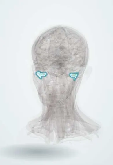
Joints and ligaments · Head and neck
TMJ capsule !
The capsule of the temporomandibular (jaw) joint is the sleeve of tissue around the joint in front of the ear; it holds the joint together and limits it.
Official name: Temporomandibular joint
Opens in the Durgical app, no sign-up needed.
What it is
The sleeve of tissue around the jaw joint, thickened on the outside.
What it does
Holds the joint together and limits how far the jaw drops back.
Key facts
- Attaches
- Margins of the articular fossa and eminence to the neck of the condyle; thickened laterally as the lateral (temporomandibular) ligament
- Function
- Encloses the TMJ; the lateral ligament stops the condyle being pushed back
Relations
- Lateral
- Parotid gland, the superficial temporal artery and the auriculotemporal nerve; temporal and zygomatic branches of the facial nerve cross just in front
- Behind
- Retrodiscal tissue, then the tympanic plate and the ear canal
- In front
- Lateral pterygoid inserting into the condylar neck, capsule and disc
- Medial
- Sphenomandibular ligament; the middle meningeal artery rising to the foramen spinosum and the chorda tympani nearby
- Contents
- Condyle of the mandible and the TMJ disc, which divides the joint into upper and lower synovial compartments
How to find it
With a fingertip just in front of the tragus, the condyle is felt moving forward under the capsule as the mouth opens; a finger in the ear canal pressing forward feels the back of the condyle.
Examination
- Palpation of the lateral pole in front of the tragus: tenderness suggests capsulitis or synovitis
- Mouth opening (distance between the incisors) and its path: limited opening or deviation suggests a joint or muscle problem
On imaging
MRI with closed- and open-mouth views shows the capsule, joint effusion and the disc; CT or cone-beam CT shows the bony surfaces; ultrasound can show an effusion.
Clinical notes
- Arthrocentesis and arthroscopy enter the upper compartment through the lateral capsule just in front of the tragus; the facial nerve branches are the main structures at risk
- Rheumatoid arthritis and juvenile idiopathic arthritis can involve the TMJ synovium, causing pain and stiffness and, in children, reduced jaw growth
- Ankylosis after trauma or infection in childhood fuses the condyle to the fossa, limiting mouth opening and jaw growth
Development
The TMJ forms later than most synovial joints, as a secondary joint between the temporal bone and the condylar cartilage; that cartilage stays a growth site through childhood, so condylar injury or ankylosis at a young age can stunt the mandible on that side.
Related conditions
Test yourself
Think of the answer, then tap the question to see it.
What does the lateral (temporomandibular) ligament of the TMJ capsule prevent?
It stops the condyle being driven back against the tympanic plate and ear canal.
Why can TMJ capsule pain be felt in the ear?
Its sensory supply is mainly from the auriculotemporal nerve (V3).
In anterior TMJ dislocation, where is the condyle and what holds it?
In front of the articular eminence, held by muscle spasm; the capsule usually stays intact.
Which structures are mainly at risk when TMJ arthrocentesis enters the capsule in front of the tragus?
The facial nerve branches.
Educational information, not medical advice. For any symptom, see a health professional.
Its names
- Official Latin (TA2)
- articulatio temporomandibularis TA2 1622
- Official Spanish (DeCS)
- Cápsula de la articulación temporomandibular DeCS D013704
- English (TA2)
- Temporomandibular joint
- Also called
- articulatio mandibularis · articulus mandibularis
Sources: Terminologia Anatomica (TA2); DeCS; Anatomy, Head and Neck, Temporomandibular Joint.
Spotted a mistake?We are always improving for the community. Tell us what is wrong and our team will review it.Report a mistake
Representative model, for educational purposes only.