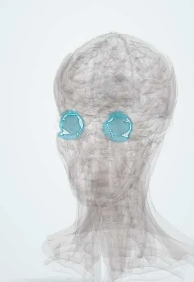
Eye fascia !
The eye fascia is a thin sheath around the eyeball with a supporting hammock below; it lets the eye turn smoothly and holds it up.
Official name: Suspensory ligament of eyeball
Opens in the Durgical app, no sign-up needed.
What it is
A thin sheath around the eyeball with a hammock underneath.
What it does
Lets the eye turn smoothly and supports it from below.
Key facts
- Attaches
- Fascial sheath (Tenon capsule) from the optic nerve to the limbus; suspensory ligament (Lockwood) slung below the eye between the lacrimal and zygomatic bones
- Function
- Socket in which the eye turns; holds the eye up if the orbital floor breaks
Relations
- Superficial
- Bulbar conjunctiva, fused to it in the last few millimetres before the limbus
- Deep
- Sclera, separated by the thin episcleral space crossed by fine connective strands
- Behind
- Blends with the dural sheath of the optic nerve; orbital fat lies behind and around it
- Medial
- Medial check ligament to the lacrimal bone, behind the posterior lacrimal crest
- Lateral
- Lateral check ligament to the orbital tubercle of the zygomatic bone
- Below
- The suspensory ligament, a hammock under the globe; in front it continues as the capsulopalpebral fascia into the lower lid, so the lower lid follows the eye on downgaze
How to find it
It is met in surgery after the bulbar conjunctiva is opened near the limbus: a whitish layer over the sclera, thicker in young people, which is opened in turn to reach the rectus muscle insertions.
On imaging
Orbital ultrasound (B-scan) in posterior scleritis shows fluid in the sub-Tenon space around the back of the globe and the optic nerve, the "T sign".
Clinical notes
- Sub-Tenon anaesthesia: a blunt cannula passed into the episcleral space spreads anaesthetic around the globe for cataract and other eye surgery, without a sharp needle behind the eye
- After removal of the eye (enucleation), an orbital implant is placed inside the sheath and the recti are attached to it, so the artificial eye can move
- In strabismus surgery the muscles are reached through the conjunctiva and the sheath; in glaucoma filtering surgery, scarring of the sheath over the drainage bleb is a main cause of failure
Related conditions
Test yourself
Think of the answer, then tap the question to see it.
What is the fascial sheath of the eyeball (Tenon capsule), and what does the space beneath it allow?
A sheath wrapping the globe from the optic nerve to near the limbus; the episcleral (sub-Tenon) space lets the eye turn like a ball in a socket.
What forms the suspensory ligament of the eye (Lockwood)?
Mainly the fused sheaths of the inferior rectus and inferior oblique, joined at each end to the check ligaments; it slings the globe from below.
How is sub-Tenon anaesthesia given for eye surgery?
A blunt cannula is passed into the episcleral space to spread anaesthetic around the globe, without a sharp needle behind the eye.
Educational information, not medical advice. For any symptom, see a health professional.
Its names
- Official Latin (TA2)
- ligamentum suspensorium bulbi TA2 6818
- Official Spanish (DeCS)
- Vaina del globo ocular
- English (TA2)
- Suspensory ligament of eyeball
- Also called
- lockwood’s ligament
Sources: Terminologia Anatomica (TA2); Anatomy, Head and Neck, Eye Extraocular Muscles; Anatomy, Head and Neck, Orbit.
Spotted a mistake?We are always improving for the community. Tell us what is wrong and our team will review it.Report a mistake
Representative model, for educational purposes only.