Orbital cellulitis !
Orbital cellulitis is an infection of the tissues around the eye inside the eye socket, usually spread from the sinuses; it can threaten sight and life.
Updated 2026-10-29
Structures involved
Tap one to see what it is and open it in 3D.
- Eye fascia
- Ethmoid cells
- Eyelids
- Optic nerve
- Cavernous sinus
- Ophthalmic veins
- Maxillary sinus
- Frontal sinus
Why it happens
What it is
Infection of the soft tissues of the orbit behind the orbital septum, usually spreading from the ethmoid sinuses. It differs from preseptal cellulitis, which stays in front of the septum, and can threaten vision and life.
Who gets it
Commoner in children and adolescents, in whom ethmoid sinusitis is the usual source. More frequent in the colder months when respiratory infections peak.
Causes
- Spread from acute sinusitis, especially ethmoid, through the thin lamina papyracea
- Trauma or orbital surgery
- Dental infection
- Spread from preseptal cellulitis, dacryocystitis or eyelid infection
- Organisms: streptococci, Staphylococcus aureus (including MRSA), Haemophilus influenzae, anaerobes; fungi (mucormycosis) in immunocompromised or diabetic patients
Risk factors
- Acute bacterial sinusitis
- Childhood and adolescence
- Recent upper respiratory infection
- Penetrating eyelid or orbital trauma
- Diabetes (especially ketoacidosis) and immunosuppression
- Incomplete Haemophilus influenzae type b vaccination
Pathophysiology
Bacteria pass from the ethmoid sinus through the thin medial orbital wall or through valveless orbital veins. Inflammation behind the septum raises orbital pressure and pushes the eye forward, limits muscle movement and can compress the optic nerve. Infection can collect as a subperiosteal or orbital abscess or spread through the ophthalmic veins to the cavernous sinus.
Classification
- Chandler classification: preseptal cellulitis, orbital cellulitis, subperiosteal abscess, orbital abscess, cavernous sinus thrombosis
Symptoms and signs
- Painful swelling and redness of the eyelids
- Pain on moving the eye and limited eye movement
- Proptosis (bulging eye)
- Double vision
- Chemosis (conjunctival swelling)
- Fever and malaise
- Reduced vision or colour vision in severe cases
Diagnosis
- Clinical
- Proptosis, painful or restricted eye movements, reduced vision or chemosis separate orbital from preseptal cellulitis. Visual acuity, colour vision, pupils and eye movements are checked repeatedly.
- Tests
- Blood count, inflammatory markers and blood cultures. Cultures of pus from drained abscesses or sinuses guide antibiotics.
- Imaging
- Contrast CT of the orbits and sinuses confirms orbital involvement, shows abscesses and the sinus source. MRI if intracranial spread or cavernous sinus thrombosis is suspected.
Differential diagnosis
- Preseptal cellulitis
- Allergic or insect-bite eyelid swelling
- Thyroid eye disease
- Idiopathic orbital inflammation
- Orbital tumour (rhabdomyosarcoma in children)
- Ruptured dermoid cyst
Goal of treatment
Control the infection quickly with antibiotics, drain abscesses when needed, protect the optic nerve and prevent intracranial spread.
Treatment
- Acute
- Hospital admission with intravenous broad-spectrum antibiotics covering streptococci, staphylococci (including MRSA where common) and anaerobes, changed to oral when clearly improving.
- Follow-up
- Frequent checks of vision, pupils, colour vision and eye movements, with joint ophthalmology and ENT care.
- Medical
- Nasal decongestants and saline irrigation for the sinus source; corticosteroids are sometimes added once infection is controlled, though evidence is limited.
- Surgical
- Drainage of a subperiosteal or orbital abscess, and endoscopic sinus surgery, when vision worsens, the abscess is large, or there is no response to antibiotics.
- Surgical
- Mucormycosis: urgent surgical debridement plus systemic antifungals and control of diabetes or immunosuppression.
Complications
- Permanent vision loss from optic nerve compression or ischaemia
- Subperiosteal or orbital abscess
- Cavernous sinus thrombosis
- Meningitis, epidural or subdural empyema, brain abscess
- Sepsis
Prognosis
With early antibiotics and timely drainage, most patients recover fully. Delay, abscess formation and fungal infection increase the risk of blindness and death.
Prevention
- Haemophilus influenzae type b and pneumococcal vaccination
- Prompt treatment of complicated bacterial sinusitis
- Good glycaemic control in diabetes
Test yourself
Think of the answer, then tap the question to see it.
Which sinus is the commonest source of orbital cellulitis?
The ethmoid sinus, via the thin lamina papyracea of the medial orbital wall.
Which findings separate orbital from preseptal cellulitis?
Proptosis, painful or limited eye movements, reduced vision and chemosis.
What imaging is done when orbital cellulitis is suspected?
Contrast CT of the orbits and sinuses.
Which classification stages orbital infection from preseptal to cavernous sinus thrombosis?
The Chandler classification.
Which life-threatening fungal infection causes orbital cellulitis in diabetic ketoacidosis?
Mucormycosis (rhino-orbital-cerebral), with black necrotic eschar and rapid spread.
Related conditions
- Acute rhinosinusitis
- Bacterial meningitis
- Chalazion and hordeolum (stye)
- Orbital floor (blowout) fracture
Educational information, not medical advice. For any symptom, see a health professional.
Sources: Orbital Cellulitis (StatPearls (NCBI Bookshelf), 2026).
Spotted a mistake?We are always improving for the community. Tell us what is wrong and our team will review it.Report a mistake
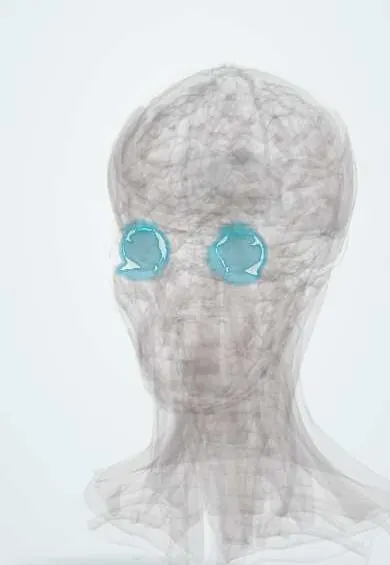
Representative model, for educational purposes only.