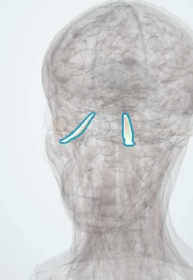
Inferior rectus !
The inferior rectus is one of the six muscles that move the eyeball, under the eye inside the eye socket; it turns the eye down and slightly in.
Official name: Inferior rectus muscle
Opens in the Durgical app, no sign-up needed.
What it is
One of the six muscles that move the eyeball, inside the eye socket.
What it does
Turns the eye down (and slightly in).
Key facts
- Origin
- Common tendinous ring (annulus of Zinn)
- Insertion
- Sclera below, ~6.5 mm behind the limbus
- Action
- Depression (best in abduction); extorsion, adduction
- Nerve
- Oculomotor nerve (III), inferior division
- Blood supply
- Ophthalmic artery (muscular branches)
Relations
- Below
- Orbital floor (maxilla), with the infraorbital nerve and vessels in their groove and canal; below the floor, the maxillary sinus
- Above
- Globe and optic nerve
- Lateral
- The nerve to the inferior oblique, which runs forward along its lateral border; the lateral rectus further out
- In front
- Near its insertion the inferior oblique crosses below it; their sheaths fuse into the suspensory (Lockwood) ligament
How to find it
Its tendon inserts about 6.5 mm behind the lower limbus and is reached through the lower fornix. From below it lies on the orbital floor, near the thin bone beside the infraorbital groove; toward the front the inferior oblique passes under it, between the muscle and the floor.
Examination
- H-pattern: the person looks out, then down; weak depression in abduction isolates the inferior rectus
- Forced duction test: under topical anaesthesia the eye is grasped and moved; resistance to moving it up means a mechanical restriction (trapped or fibrotic inferior rectus), not a nerve palsy
On imaging
Coronal CT of the orbits is the key view. In a blowout fracture, look for orbital fat or muscle hanging into the maxillary sinus (the "teardrop" sign) and a rounded or trapped inferior rectus. In thyroid eye disease the muscle bellies are enlarged while the tendons are spared.
Common problems
- Sixth-nerve palsy: the eye cannot turn out, causing side-by-side double vision.
- Strabismus (squint) when the eye muscles are unbalanced.
How it is diagnosed
Eye movement and alignment tests; scans if a nerve palsy is new.
Treatment
- Prisms or patching; botulinum toxin; eye muscle surgery to rebalance the muscles.
Clinical notes
- In children a trapdoor floor fracture can grip the muscle with almost no bruising ("white-eyed blowout"); pain on upgaze, nausea, vomiting and a slow heart rate (oculocardiac reflex) make it a surgical emergency
- Numbness of the cheek, upper lip and upper teeth after a floor fracture shows injury to the infraorbital nerve
- Thyroid eye disease: enlarged muscles push the eye forward (exophthalmos) and can compress the optic nerve at a crowded orbital apex
Development
Arises from the shared head-mesoderm mass of the oculomotor muscles near the optic stalk; congenital fibrosis of the extraocular muscles mainly stiffens this group, often leaving the eyes held down with ptosis.
Memory aid
Muscles enlarged in thyroid eye disease, most to least often: "I'M SLOw" (Inferior, Medial, Superior, Lateral, Obliques).
Related conditions
- Graves disease (hyperthyroidism)
- Orbital floor (blowout) fracture
- Thyroid eye disease (Graves orbitopathy)
Test yourself
Think of the answer, then tap the question to see it.
Which extraocular muscle is most often trapped in an orbital floor (blowout) fracture, and what is the sign?
The inferior rectus: upgaze is limited and the person sees double looking up.
What is a «white-eyed blowout» in a child, and why is it an emergency?
A trapdoor floor fracture gripping the inferior rectus with little bruising; pain on upgaze, nausea and a slow heart rate (oculocardiac reflex) need urgent surgery.
Which muscle is usually the first and most enlarged in thyroid eye disease?
The inferior rectus; when tight it holds the eye down and limits upgaze.
What does the forced duction test tell in limited upgaze?
Resistance to moving the eye up means a mechanical restriction (trapped or fibrotic inferior rectus), not a nerve palsy.
What does cheek, upper lip and upper teeth numbness after a floor fracture indicate?
Injury to the infraorbital nerve, which runs in the orbital floor below the inferior rectus.
Educational information, not medical advice. For any symptom, see a health professional.
Its names
- Official Latin (TA2)
- musculus rectus inferior TA2 2043
- Official Spanish (DeCS)
- Músculo recto inferior DeCS D009801
- English (TA2)
- Inferior rectus muscle
- Also called
- musculus rectus inferior bulbi
Related
Sources: Terminologia Anatomica (TA2); DeCS; Anatomy, Head and Neck: Eye Inferior Rectus Muscle; Anatomy, Head and Neck, Eye Extraocular Muscles; Anatomy of the Human Body (20th ed.): The Accessory Organs of the Eye (Lea & Febiger (Gray's Anatomy, 1918), 1918) · adapted, Public domain; Anatomy of the Human Body (20th ed.): Development of the Muscles (Lea & Febiger (Gray's Anatomy, 1918), 1918) · adapted, Public domain.
Spotted a mistake?We are always improving for the community. Tell us what is wrong and our team will review it.Report a mistake
Representative model, for educational purposes only.