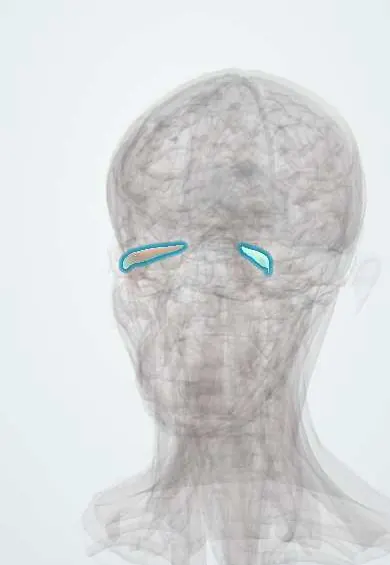
Lateral rectus !
The lateral rectus is one of the six muscles that move the eyeball, on its outer side; it turns the eye outward and is supplied by the sixth cranial nerve.
Official name: Lateral rectus muscle
Opens in the Durgical app, no sign-up needed.
What it is
One of the six muscles that move the eyeball, inside the eye socket.
What it does
Turns the eye outward; supplied by nerve VI.
Key facts
- Origin
- Common tendinous ring (annulus of Zinn)
- Insertion
- Sclera laterally, ~6.9 mm behind the limbus
- Action
- Abduction
- Nerve
- Abducens nerve (VI)
- Blood supply
- Ophthalmic artery (muscular branches)
Relations
- Lateral
- Lateral orbital wall (zygomatic bone and greater wing of the sphenoid); at the front, the lacrimal gland above it
- Medial
- Ciliary ganglion and optic nerve; the abducens nerve enters its inner (ocular) surface
- Above
- Lacrimal artery and lacrimal nerve running along its upper border to the lacrimal gland
- Below
- Orbital floor; the inferior oblique inserts on the globe beneath its back end
How to find it
Its tendon inserts about 6.9 mm behind the lateral limbus and is reached through a temporal conjunctival incision. In dissection it follows the lateral orbital wall; at the apex its two heads straddle the superior orbital fissure, and in front the lacrimal gland rests on its tendon.
Examination
- H-pattern: the person looks straight out to the side; an eye that lags or fails to pass the midline points to the lateral rectus or the abducens nerve (VI)
- Head posture: a person with a sixth-nerve palsy often turns the face toward the weak side to keep single vision
On imaging
Axial CT or MRI shows it along the lateral orbital wall to the globe; coronal images compare its size with the other recti. In thyroid eye disease it is usually the last rectus to enlarge. In a sixth nerve palsy, imaging targets the nerve's long path: MRI of the brain and skull base (clivus, petrous apex, cavernous sinus), with venography if raised intracranial pressure is suspected.
Common problems
- Sixth-nerve palsy: the eye cannot turn out, causing side-by-side double vision.
- Strabismus (squint) when the eye muscles are unbalanced.
How it is diagnosed
Eye movement and alignment tests; scans if a nerve palsy is new.
Treatment
- Prisms or patching; botulinum toxin; eye muscle surgery to rebalance the muscles.
Clinical notes
- Causes of a sixth-nerve palsy: microvascular disease in adults, raised intracranial pressure, cavernous sinus lesions, tumours of the clivus and petrous apex infection (Gradenigo syndrome); in children, also brainstem tumours
- Inside the cavernous sinus the abducens nerve runs free beside the internal carotid artery, so it is often the first nerve affected by a carotid aneurysm or cavernous sinus thrombosis
- Duane syndrome (congenital): the abducens nucleus is missing and a branch of the oculomotor nerve reaches the lateral rectus; abduction is limited, and on adduction the globe retracts and the lid opening narrows
- Exotropia (an eye turned out) in children is often intermittent, worse when tired or in bright light; surgery may weaken the lateral rectus
Development
Develops from head mesoderm near the optic stalk but, unlike its neighbours, is supplied by the abducens nerve; when that nerve fails to form, the muscle can be innervated by the oculomotor nerve instead (the basis of Duane syndrome).
Memory aid
"LR6 SO4, rest 3": lateral rectus → abducens (VI); superior oblique → trochlear (IV); all the others → oculomotor (III).
Related conditions
Test yourself
Think of the answer, then tap the question to see it.
Which nerve supplies the lateral rectus, and what does its palsy cause?
The abducens nerve (VI): the eye turns in and cannot abduct, with side-by-side double vision worse at distance and toward the weak side.
Why is a sixth-nerve palsy called a false localising sign?
The abducens nerve has a long intracranial course, so raised intracranial pressure can stretch it, wherever the lesion is.
Why is the abducens nerve often the first affected in cavernous sinus thrombosis or a carotid aneurysm?
It runs free in the cavernous sinus beside the internal carotid artery.
What is Duane syndrome?
Congenital absence of the abducens nucleus, with an oculomotor branch reaching the lateral rectus: limited abduction, and globe retraction with lid narrowing on adduction.
Educational information, not medical advice. For any symptom, see a health professional.
Its names
- Official Latin (TA2)
- musculus rectus lateralis bulbi oculi TA2 2045
- Official Spanish (DeCS)
- Músculo recto lateral DeCS D009801
- English (TA2)
- Lateral rectus muscle
- Also called
- external rectus · musculus rectus termporalis bulbi
Related
Sources: Terminologia Anatomica (TA2); DeCS; Anatomy, Head and Neck: Eye Lateral Rectus Muscle; Anatomy, Head and Neck, Eye Extraocular Muscles; Anatomy of the Human Body (20th ed.): The Accessory Organs of the Eye (Lea & Febiger (Gray's Anatomy, 1918), 1918) · adapted, Public domain; Anatomy of the Human Body (20th ed.): Development of the Muscles (Lea & Febiger (Gray's Anatomy, 1918), 1918) · adapted, Public domain.
Spotted a mistake?We are always improving for the community. Tell us what is wrong and our team will review it.Report a mistake
Representative model, for educational purposes only.