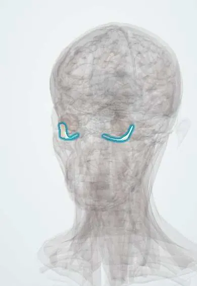
Inferior oblique !
The inferior oblique is one of the six muscles that move the eyeball, running under the eye; it turns the eye up and out and rotates it outward.
Official name: Inferior oblique muscle
Opens in the Durgical app, no sign-up needed.
What it is
One of the six muscles that move the eyeball, inside the eye socket.
What it does
Turns the eye up and out and rotates it outward.
Key facts
- Origin
- Orbital surface of the maxilla at the front of the floor, just lateral to the opening of the nasolacrimal canal
- Insertion
- Posterolateral sclera below the lateral rectus
- Action
- Extorsion; elevation (best in adduction); abduction
- Nerve
- Oculomotor nerve (III), inferior division
- Blood supply
- Infraorbital and ophthalmic arteries
Relations
- Course
- From the front of the orbital floor it runs backward and outward beneath the inferior rectus, then curves up under the lateral rectus to the posterolateral globe
- Below
- Orbital floor (maxilla) and, below it, the maxillary sinus
- Above
- Inferior rectus, which it crosses; their sheaths form the suspensory ligament
- Lateral
- Lateral rectus, under whose back end it inserts
- Medial
- Nasolacrimal duct in its bony canal, just medial to the origin
How to find it
It cannot be felt. It starts on the orbital floor just behind the lower rim, lateral to the opening of the nasolacrimal canal, then runs back and out beneath the inferior rectus to the back of the globe. Surgeons reach it through the lower temporal fornix, hooking its belly lateral to the inferior rectus.
Examination
- H-pattern: the person looks in toward the nose, then up; poor elevation in adduction isolates the inferior oblique
- Inferior oblique overaction: the adducting eye shoots up on side gaze; common in childhood strabismus and with superior oblique palsy
On imaging
Hard to see on axial images because it runs obliquely under the globe; coronal and sagittal CT or MRI show it along the front of the orbital floor, lateral to the nasolacrimal canal. In an orbital floor (blowout) fracture, coronal CT shows the defect and any trapped tissue; the inferior rectus is the muscle usually caught, and the inferior oblique origin lies just in front of the fracture zone.
Common problems
- Sixth-nerve palsy: the eye cannot turn out, causing side-by-side double vision.
- Strabismus (squint) when the eye muscles are unbalanced.
How it is diagnosed
Eye movement and alignment tests; scans if a nerve palsy is new.
Treatment
- Prisms or patching; botulinum toxin; eye muscle surgery to rebalance the muscles.
Clinical notes
- Overacting inferior obliques give a V pattern (the eyes diverge more on upgaze); weakening the muscle is a common strabismus operation
- Lower lid and orbital floor surgery work close to its origin; injury causes vertical double vision
- The inferotemporal vortex vein lies next to its insertion and is at risk during surgery on the muscle
Development
Belongs to the common mass of head mesoderm near the optic stalk from which all the muscles of the oculomotor nerve develop.
Memory aid
The obliques do the opposite of their name for up and down: the superior oblique depresses and the inferior oblique elevates; both abduct.
Related conditions
Test yourself
Think of the answer, then tap the question to see it.
What is unique about the origin of the inferior oblique among the extraocular muscles?
It is the only one not arising at the orbital apex: it starts at the front of the orbital floor, beside the nasolacrimal canal.
In which gaze direction is the inferior oblique tested?
Looking up and in: with the eye adducted it is the main elevator.
Which nerve supplies the inferior oblique, and what extra fibres does it carry?
The inferior division of the oculomotor nerve (III); its nerve carries the parasympathetic fibres to the ciliary ganglion.
What pattern do overacting inferior obliques produce?
A V pattern: the eyes diverge more on upgaze; the adducting eye shoots up on side gaze.
Educational information, not medical advice. For any symptom, see a health professional.
Its names
- Official Latin (TA2)
- musculus obliquus inferior bulbi oculi TA2 2051
- Official Spanish (DeCS)
- Músculo oblicuo inferior DeCS D009801
- English (TA2)
- Inferior oblique muscle
- Also called
- musculus obliquus inferior bulbi · Oblicuo menor
Related
Sources: Terminologia Anatomica (TA2); DeCS; Anatomy, Head and Neck: Inferior Oblique Muscle; Anatomy, Head and Neck, Eye Extraocular Muscles; Anatomy of the Human Body (20th ed.): The Accessory Organs of the Eye (Lea & Febiger (Gray's Anatomy, 1918), 1918) · adapted, Public domain; Anatomy of the Human Body (20th ed.): Development of the Muscles (Lea & Febiger (Gray's Anatomy, 1918), 1918) · adapted, Public domain.
Spotted a mistake?We are always improving for the community. Tell us what is wrong and our team will review it.Report a mistake
Representative model, for educational purposes only.