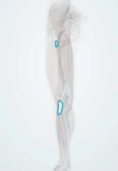
Vastus medialis tendon !
The vastus medialis tendon is the part of the quadriceps tendon from the inner thigh muscle; it pulls on the inner edge of the kneecap.
Official name: Tendon of vastus medialis
Opens in the Durgical app, no sign-up needed.
What it is
The part of the quadriceps tendon that comes from the inner thigh muscle.
What it does
Transmits the force of the quadriceps to straighten the knee.
Key facts
- Attaches
- Middle layer of the quadriceps tendon to the medial base and border of the patella; oblique lower fibres (VMO) to the upper medial patella; expansion to the medial retinaculum
- Function
- Extends the knee; the oblique fibres resist lateral patellar tracking, most in the last degrees of extension
- Tight in
- Knee flexion
Relations
- Deep
- MPFL and the knee joint capsule medially; the vastus intermedius tendon higher up
- Lateral
- Vastus lateralis and rectus femoris tendons, which it meets at the base of the patella
- Medial
- Adductor magnus tendon and the medial intermuscular septum, from which the oblique fibres arise
- Superficial
- Fascia and skin; the teardrop bulge above the inner patella is the VMO
How to find it
With the knee fully straightened and the thigh tightened, the teardrop bulge above and inside the patella is the VMO; its tendinous end is felt where the bulge merges into the upper inner border of the patella.
Examination
- Patellar apprehension test: the examiner pushes the patella laterally with the knee slightly bent; guarding or fear of dislocation suggests weak medial restraints (MPFL, VMO)
On imaging
MRI shows it best: on axial images the lowest VMO fibres reach the upper medial patella, with the MPFL just deep to them. After a lateral patellar dislocation, look for oedema or tearing of these fibres with an MPFL tear, and bone bruises on the medial patella and the lateral femoral condyle. Lateral and axial (skyline) radiographs look for a small avulsed fragment and patellar position.
Common problems
- Tendinitis from repeated jumping or kneeling.
- Rupture, usually in older adults; the leg cannot be straightened.
How it is diagnosed
Physical exam; ultrasound or MRI.
Treatment
- Tendinitis: rest, physical therapy.
- Complete tear: surgical repair, usually soon after the injury.
- Part of this tendon can also be used as an ACL graft.
Clinical notes
- The medial parapatellar approach for knee replacement divides the junction of this tendon with the quadriceps tendon; the subvastus and midvastus approaches aim to spare it
- After a lateral patellar dislocation, MRI often shows oedema or detachment of the lowest VMO fibres next to a torn MPFL
Test yourself
Think of the answer, then tap the question to see it.
What are the vastus medialis obliquus (VMO) fibres taught to do?
Act as the main active brake on lateral patellar displacement, most in the last degrees of extension (their real strength there is debated).
Which ligament lies just deep to the lowest VMO fibres?
The medial patellofemoral ligament (MPFL), partly joined to them; both are often injured in a lateral patellar dislocation.
Which knee replacement approaches aim to spare the vastus medialis tendon?
The subvastus and midvastus approaches; the medial parapatellar approach divides its junction with the quadriceps tendon.
Educational information, not medical advice. For any symptom, see a health professional.
Its names
- Official Latin (TA2)
- musculus vastus medialis TA2 2620
- Official Spanish (DeCS)
- Tendón del vasto medial DeCS D013710
- English (TA2)
- Tendon of vastus medialis
- Also called
- musculus vastus tibialis
Related
Sources: Terminologia Anatomica (TA2); DeCS; Anatomy, Bony Pelvis and Lower Limb: Thigh Quadriceps Muscle.
Spotted a mistake?We are always improving for the community. Tell us what is wrong and our team will review it.Report a mistake
Representative model, for educational purposes only.