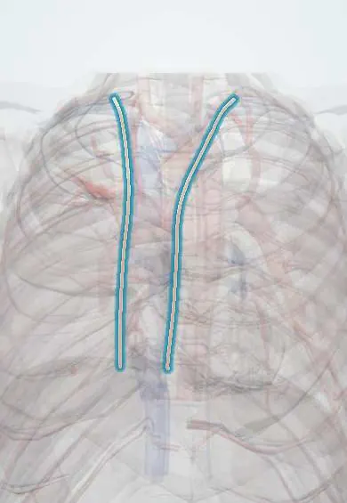
Internal thoracic artery !
The internal thoracic artery runs down inside the front chest wall beside the breastbone; it supplies the chest wall and breast and is used in bypass surgery.
Opens in the Durgical app, no sign-up needed.
What it is
An artery running down the inside of the front chest wall, about 1-2 cm beside the sternum (also called the internal mammary artery).
What it does
Supplies the chest wall and breast. It rarely gets clogged, which makes it the best bypass graft.
Key facts
- Origin
- First part of subclavian artery; descends about 1 cm lateral to sternum behind costal cartilages
- Branches
- Pericardiacophrenic (with the phrenic nerve); mediastinal and thymic; anterior intercostals (two per space, spaces 1–6); perforating (2nd–4th enlarged to the breast); ends in 6th space as musculophrenic (spaces 7–9) and superior epigastric
- Supplies
- Anterior chest wall, breast, pericardium, diaphragm, upper anterior abdominal wall; common coronary bypass graft
Relations
- Course
- From the first part of the subclavian artery it descends behind the medial end of the clavicle and the 1st costal cartilage, is crossed by the phrenic nerve, then runs straight down behind the upper six costal cartilages and divides in the 6th intercostal space
- In front
- Costal cartilages and the front ends of the internal intercostal muscles; pectoralis major further out
- Behind
- Endothoracic fascia and parietal pleura above; the transversus thoracis muscle below the 3rd costal cartilage
- Medial
- Sternal edge; the internal thoracic veins, which merge into a single vein on its medial side higher up, and the parasternal lymph nodes
How to find it
Not palpable. Its line is projected about a finger-breadth (1–2 cm) lateral to the sternal edge from the 1st to the 6th costal cartilage; parasternal punctures are planned away from this line, and ultrasound shows it as a pulsating vessel deep to the cartilages.
On imaging
On contrast CT it is a small vessel a finger-breadth from the sternal edge, behind the costal cartilages, between its veins. Before bypass, CT or angiography checks it and the subclavian for narrowing; afterwards, CT angiography shows if the graft is open. In coarctation it is enlarged on CT, and rib notching from the intercostal collaterals may show on radiographs.
Common problems
- Rarely diseased.
Treatment
- In bypass surgery the left internal mammary artery is freed and joined to the LAD; it often stays open for decades.
Clinical notes
- Harvested through a median sternotomy as a pedicle with its veins and fat, or skeletonized; taking both arteries reduces sternal blood supply and raises wound risk, especially in diabetes
- A narrowing of the subclavian artery before its origin can steal flow from a coronary graft (coronary-subclavian steal), with angina on arm effort
- Parasternal stab wounds can cut it and cause a large haemothorax
- Used as the recipient artery in free-flap breast reconstruction
Anatomical variants
- Origin and branching vary: usually from the first part of the subclavian artery, occasionally from the thyrocervical trunk or a shared trunk
- A lateral costal branch running down the inner chest wall behind the ribs is present in some people
Related conditions
Test yourself
Think of the answer, then tap the question to see it.
From which artery does the internal thoracic artery arise, and where does it run in the chest?
From the first part of the subclavian artery; it descends about a finger-breadth (1–2 cm) lateral to the sternal edge, behind the upper six costal cartilages.
What are the two terminal branches of the internal thoracic artery, and where does it divide?
The musculophrenic and superior epigastric arteries, in the 6th intercostal space.
Why is the left internal thoracic artery the conduit of choice in coronary bypass surgery?
Grafted to the left anterior descending artery, it rarely clogs and has excellent long-term patency, often staying open for decades.
What is coronary-subclavian steal?
A narrowing of the subclavian artery before the origin of an internal thoracic artery graft steals flow from the coronary graft, causing angina on arm effort.
How does the internal thoracic artery provide a collateral route around an aortic coarctation?
Its superior epigastric branch joins the inferior epigastric artery in the rectus sheath.
Educational information, not medical advice. For any symptom, see a health professional.
Its names
- Official Latin (TA2)
- arteria thoracica interna; rami intercostales anteriores arteriae thoracicae internae TA2 4576
- Official Spanish (DeCS)
- Arterias mamarias DeCS D008323
- English (TA2)
- Internal thoracic artery
- Also called
- internal mammary artery · mammary arteries · LIMA · arteria mammaria interna
Related
Sources: Terminologia Anatomica (TA2); DeCS; Anatomy, Thorax, Internal Mammary (Internal Thoracic) Arteries; Tratado de anatomía humana (9.ª ed.), tomo II, Libro IV Angiología, arterias subclavia, axilar, humeral, radial y cubital, arcos palmares y vías anastomóticas del miembro superior.
Spotted a mistake?We are always improving for the community. Tell us what is wrong and our team will review it.Report a mistake
Representative model, for educational purposes only.