
Angle-recession glaucoma
A blunt blow to the eye can tear the drainage angle. Years later, scarring of the drain can raise pressure and cause glaucoma in that eye.
What it is
A unilateral secondary open-angle glaucoma after blunt trauma that tore between the longitudinal and circular muscles of the ciliary body. Gonioscopy shows an irregularly widened ciliary body band. Glaucoma may appear months to decades later, and the risk rises with the extent of the recession.
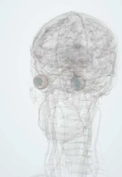
See it in 3D
Who gets it
Angle recession is found in most eyes with traumatic hyphaema, but only a small fraction develop glaucoma; young men are most often affected because of their higher trauma rates.
Causes
- Blunt ocular trauma (balls, fists, projectiles, airbags)
Risk factors
- Recession involving more than about 180 degrees of the angle
- Traumatic hyphaema
- Pre-existing tendency to open-angle glaucoma (the fellow eye is also at risk)
Pathophysiology
Sudden compression of the globe forces aqueous into the angle and splits the ciliary muscle. The meshwork is damaged at the same time and later scars or is covered by a membrane, which reduces outflow and slowly raises pressure.
Symptoms and signs
- None in the early stages
- Late peripheral field loss in the injured eye
Diagnosis
- Clinical
- Gonioscopy compared with the fellow eye: widened ciliary body band, torn iris processes, a whiter scleral spur; other trauma signs such as iris sphincter tears, iridodialysis or phacodonesis.
- Tests
- Tonometry, perimetry.
- Imaging
- Ultrasound biomicroscopy shows the recessed angle; OCT of the nerve fibre layer.
Differential diagnosis
- Primary open-angle glaucoma (bilateral)
- Cyclodialysis cleft (low pressure)
- Other secondary glaucomas after trauma (lens-induced, ghost cell, hyphaema)
One-sided glaucoma after blunt trauma
| Feature | Angle-recession glaucoma | Cyclodialysis cleft | Hyphaema-related glaucoma | Primary open-angle glaucoma |
|---|---|---|---|---|
| Timing after injury | Months to decades | Days to weeks | Days to weeks | Unrelated to injury |
| Pressure | High | Low (hypotony) | High | High |
| Gonioscopy | Irregularly widened ciliary body band | Gap between scleral spur and ciliary body | Red cells or khaki ghost cells in the chamber and angle | Normal open angle |
| Eyes affected | Injured eye; fellow eye at some risk | Injured eye | Injured eye | Both, often asymmetric |
| Laser trabeculoplasty | Works poorly | Not used | Not used | Effective |
Goal of treatment
Lower pressure enough to protect the optic nerve, and detect late glaucoma by lifelong follow-up.
Treatment
- Medical
- Aqueous suppressants and prostaglandin analogues; miotics may worsen it and are avoided.
- Surgical
- Trabeculectomy with antimetabolite or a glaucoma drainage device when drops are not enough; laser trabeculoplasty works poorly.
- Follow-up
- Lifelong yearly pressure checks of both eyes after significant blunt trauma.
Complications
- Glaucomatous optic atrophy
- Glaucoma in the fellow eye
Common traps
- Glaucoma after an eye injury appears within weeks or not at all: angle-recession glaucoma can start months to decades later, so significant blunt trauma needs lifelong pressure checks
- The split ciliary muscle is what blocks outflow: the tear marks the force of the blow; pressure rises because the meshwork injured at the same time scars or is covered by a membrane
- Only the injured eye is at risk: the fellow eye has a higher-than-average risk of open-angle glaucoma, so both eyes are followed
- Pilocarpine helps, as in some other glaucomas: miotics can worsen angle-recession glaucoma and are avoided
- A widened ciliary body band is obvious on its own: band width varies between people, so the suspect area is compared with the same area of the fellow eye
Prognosis
Controllable when detected, but often found late because it is painless and may start decades after injury.
Prevention
- Protective eyewear for sports and work
Test yourself
Think of the answer, then tap the question to see it.
A 40-year-old man has pressures of 32 mmHg right and 16 mmHg left, open angles, and recalls a squash-ball injury to the right eye 20 years ago. What do you look for on gonioscopy?
An irregularly widened ciliary body band in the right eye compared with the left, which points to angle-recession glaucoma.
Why does the pressure rise years after the blow rather than straight away?
The meshwork damaged by the blow slowly scars or is covered by a membrane, gradually reducing outflow.
Which extent of recession most raises the risk of glaucoma, and why are both eyes followed?
Recession over about 180 degrees of the angle; the fellow eye also has a higher-than-average risk of open-angle glaucoma.
Why is laser trabeculoplasty a poor choice in angle-recession glaucoma?
The scarred meshwork responds poorly to laser, so drops come first, then filtering surgery or a drainage device.
Connections
Educational information, not medical advice. For any symptom, see a health professional.
Sources: Traumatic Glaucoma (StatPearls Publishing (NCBI Bookshelf), 2026); Angle Recession Glaucoma (EyeWiki (American Academy of Ophthalmology), 2025).
Spotted a mistake?We are always improving for the community. Tell us what is wrong and our team will review it.Report a mistake
Representative model, for educational purposes only.