
Optic atrophy
A pale optic disc from lost optic nerve fibres. It is a sign, not a diagnosis: find the cause (pressure, glaucoma, inflammation, toxins, genes).
What it is
Optic atrophy is the end stage of damage to retinal ganglion cell axons, seen as a pale optic disc with loss of vision, colour vision and visual field. It follows many diseases: compression, glaucoma, ischaemia, optic neuritis, raised intracranial pressure, toxins, nutritional deficiency, trauma and inherited optic neuropathies.
Who gets it
Common in neuro-ophthalmology clinics; the leading causes vary by age, with compressive, ischaemic and glaucomatous causes in adults and tumours, hydrocephalus and hereditary disease in children.
Causes
- Compression: pituitary adenoma, meningioma, craniopharyngioma, aneurysm, thyroid eye disease
- Glaucoma (cupping with pallor of the remaining rim)
- Ischaemic optic neuropathy (arteritic and non-arteritic)
- Demyelinating or inflammatory optic neuritis
- Long-standing papilloedema (secondary atrophy)
- Toxic and nutritional: methanol, ethambutol, tobacco-alcohol, vitamin B12 or folate deficiency
- Hereditary: Leber hereditary optic neuropathy (mtDNA), dominant optic atrophy (OPA1)
- Trauma, radiation, retinal disease (consecutive atrophy)
Risk factors
- Glaucoma or raised intracranial pressure
- Vascular risk factors and giant cell arteritis
- Multiple sclerosis
- Malnutrition, alcohol use or exposure to methanol or ethambutol
- Family history of optic neuropathy
Pathophysiology
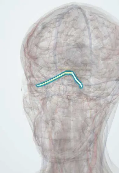
Injury anywhere from the retina to the lateral geniculate body causes degeneration of ganglion cell axons (anterograde or retrograde). The disc loses its capillaries and nerve fibre layer and turns pale. The pattern of pallor hints at the cause: temporal pallor in toxic, nutritional and hereditary disease (papillomacular bundle), band or 'bow-tie' pallor from chiasmal or optic tract lesions, and sectoral pallor after ischaemia.
Classification
- Primary (simple): flat, sharply outlined white disc, as after compression or neuritis
- Secondary: grey, blurred disc after papilloedema or papillitis, with gliosis
- Consecutive: waxy pallor from retinal disease such as retinitis pigmentosa
- Glaucomatous: pallor with enlarged cupping
- By pattern: temporal, sectoral, band (bow-tie) or diffuse
Symptoms and signs
- Gradual or previous loss of vision in one or both eyes
- Dull or washed-out colours
- Visual field defects (central scotoma, altitudinal, bitemporal)
- Reduced contrast and brightness in the affected eye
Diagnosis
- Clinical
- Visual acuity, colour vision, pupils (relative afferent pupillary defect), visual fields and fundus examination of disc colour, cup and pattern of pallor.
- Tests
- Optical coherence tomography of the nerve fibre and ganglion cell layers, visual evoked potentials, B12, folate, syphilis serology, inflammatory markers; genetic testing for LHON or OPA1 when suspected.
- Imaging
- MRI of the brain and orbits with contrast for any unexplained optic atrophy, looking for compression, demyelination or infiltration.
Differential diagnosis
- Myopic or tilted disc
- Optic nerve hypoplasia
- Physiologically large cup
- Disc pallor after cataract removal (pseudo-pallor)
Pale disc: true optic atrophy vs its look-alikes
| Feature | Optic atrophy (non-glaucomatous) | Glaucomatous atrophy | Optic nerve hypoplasia | Myopic or tilted disc |
|---|---|---|---|---|
| Disc size | Normal | Normal | Small, often with a double-ring sign | Normal or oval, often with peripapillary atrophy |
| Rim colour | Pale, out of proportion to any cupping | Remaining rim fairly pink; pallor mainly in the enlarged cup | Grey or pale but small | May look pale from the tilt without true fibre loss |
| Cup | Normal or mildly enlarged | Enlarged, often vertically, with rim notching | Small or absent | Shallow and hard to judge |
| Course | Acquired after an injury that has already cost vision | Slow progression linked to optic nerve damage from pressure | Congenital and stable | Stable |
| Key next step | MRI of brain and orbits with contrast if unexplained | Intraocular pressure, gonioscopy, visual fields, OCT | Check midline brain and pituitary (septo-optic dysplasia) | Refraction and serial OCT to confirm stability |
Goal of treatment
Find and treat the cause to protect the remaining fibres; lost axons do not regenerate.
Treatment
- Surgical
- Decompression of a compressive lesion (pituitary or orbital tumour) or shunting for hydrocephalus or raised pressure.
- Medical
- Treat the cause: lower intraocular pressure in glaucoma, corticosteroids for giant cell arteritis, stop ethambutol, replace B12 or folate.
- Rehabilitation
- Low-vision aids and visual rehabilitation for permanent loss.
- Follow-up
- Serial visual fields and optical coherence tomography to detect progression.
Complications
- Permanent loss of vision
- Legal blindness when bilateral
- Progression if the cause is missed
Common traps
- A normal-looking disc rules out optic nerve damage: after an injury behind the eye, axons die back slowly and pallor takes about 4 to 6 weeks to appear, so early retrobulbar damage shows a normal disc
- Optic atrophy is a final diagnosis: it is an end-stage sign of many diseases, and unexplained atrophy needs MRI of the brain and orbits with contrast to look for compression
- Any pale disc with a big cup is glaucoma: in glaucoma the cup enlarges while the remaining rim stays fairly pink; rim pallor out of proportion to cupping points to compression or another non-glaucomatous neuropathy
- Temporal pallor means multiple sclerosis: it marks loss of the papillomacular bundle, which also happens in toxic, nutritional (ethambutol, methanol, B12 deficiency) and hereditary neuropathies such as LHON and OPA1
- Bow-tie atrophy comes from a retinal lesion: horizontal band pallor reflects loss of nasal retinal fibres, which cross at the chiasm, so it localises to the chiasm or optic tract
Prognosis
Lost vision rarely returns, but early treatment of compression or toxicity can stop progression and sometimes recover part of it.
Prevention
- Early treatment of glaucoma and raised intracranial pressure
- Monitoring of vision on ethambutol
- Avoiding methanol-adulterated drinks
Test yourself
Think of the answer, then tap the question to see it.
A 50-year-old has horizontal band pallor of both discs and bitemporal field loss. Where is the lesion and what comes next?
The chiasm: crossing nasal fibres are lost, giving bow-tie pallor. MRI of the pituitary region with contrast and endocrine tests, looking for a pituitary adenoma or another mass.
Why does the disc look normal in the first weeks after a retrobulbar optic neuritis or optic nerve trauma?
Axons die back slowly from the injury site; loss of fibres and disc capillaries, and so pallor, takes about 4 to 6 weeks to show.
A patient on ethambutol for tuberculosis notices dull colours and blurred central vision. What pattern may follow and what should be done?
Temporal pallor with central scotomas from papillomacular bundle loss. Stop the drug promptly with the treating team; early withdrawal can halt or partly reverse the loss.
On fundus examination, what separates glaucomatous atrophy from compressive atrophy?
In glaucoma the cup enlarges and the remaining rim stays relatively pink; in compression the rim is pale out of proportion to any cupping.
A young man loses central vision in one eye, then the other within weeks; his maternal uncle had the same. Which diagnosis and which test?
Leber hereditary optic neuropathy, a mitochondrial DNA disease: test for the common mtDNA mutations. It leaves temporal or diffuse atrophy.
Why it happens
In neurology
Connections
Educational information, not medical advice. For any symptom, see a health professional.
Sources: Optic Atrophy (StatPearls Publishing (NCBI Bookshelf), 2024).
Spotted a mistake?We are always improving for the community. Tell us what is wrong and our team will review it.Report a mistake
Representative model, for educational purposes only.