Conditions · Hematology and oncology
Hereditary spherocytosis !
An inherited defect in the red cell membrane makes the cells small and round, so the spleen destroys them early, causing anaemia, jaundice and gallstones.
Updated 2026-11-01
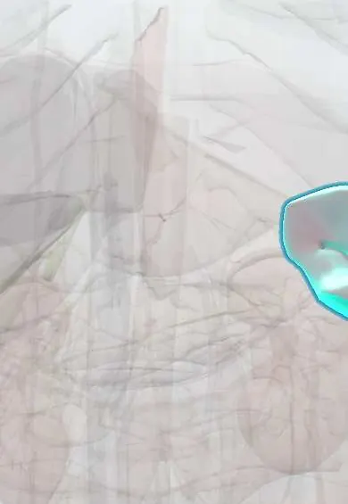
Structures involved
Tap one to see what it is and open it in 3D.
In the clinical exam
Why it happens
- Mendelian inheritance: autosomal dominant, recessive and X-linked
- The red cell life cycle and the spleen
- Membrane structure
- Lymph node and spleen under the microscope
What it is
The most common inherited red cell membrane disorder in people of European descent. Defects in proteins that anchor the membrane to its inner skeleton (ankyrin, spectrin, band 3, protein 4.2) make the cell lose surface area and become a rigid sphere that cannot squeeze through the spleen, where it is destroyed.
Who gets it
Affects roughly 1 in 2,000 people of Northern European ancestry and is found worldwide, including in Latin America, where it is a frequent cause of hereditary haemolytic anaemia alongside sickle cell disease, thalassaemias and G6PD deficiency. Severity ranges from silent carriers to transfusion-dependent infants.
Causes
- Mutations in genes for red cell membrane proteins: ANK1 (ankyrin, most common), SPTB and SPTA1 (spectrin), SLC4A1 (band 3), EPB42 (protein 4.2)
- About three quarters of cases are autosomal dominant; the rest are recessive or new mutations
Risk factors
- Family history of anaemia, jaundice, gallstones or splenectomy at a young age
- Northern European ancestry (though it occurs in all populations)
Pathophysiology
Weak vertical links between the lipid bilayer and the spectrin skeleton let small pieces of membrane bud off. The cell loses surface area relative to its volume, becomes spherical, less deformable and more concentrated (high MCHC). In the narrow passages of the splenic red pulp these cells are trapped, conditioned and engulfed by macrophages: extravascular haemolysis that enlarges the spleen and raises bilirubin. The marrow compensates with reticulocytosis, so any pause in red cell production (parvovirus B19) causes a sudden fall in haemoglobin.
Classification
- Trait or mild: compensated haemolysis, often found in adulthood
- Moderate: the most common form, anaemia and jaundice in childhood
- Severe: transfusion-dependent anaemia, usually recessive
Symptoms and signs
- Neonatal jaundice, sometimes needing phototherapy or exchange transfusion
- Pallor and tiredness from anaemia
- Intermittent jaundice, often worse during infections
- Enlarged spleen
- Biliary colic from pigment gallstones, even in childhood
Diagnosis
- Clinical
- Anaemia, jaundice and splenomegaly in a child, with a family history of the same; examine the spleen and ask about gallstones and splenectomy in relatives.
- Tests
- Complete blood count with high MCHC and reticulocytosis; peripheral smear with spherocytes lacking central pallor; haemolysis markers (high LDH and unconjugated bilirubin, low haptoglobin); negative direct antiglobulin (Coombs) test to rule out autoimmune haemolysis. Confirmation with the eosin-5-maleimide (EMA) binding test by flow cytometry or the osmotic fragility test; genetic testing in unclear cases.
- Imaging
- Abdominal ultrasound to measure the spleen and look for gallstones, especially before planned splenectomy.
Differential diagnosis
- Warm autoimmune haemolytic anaemia (Coombs positive)
- G6PD deficiency and other enzyme defects
- Other membrane disorders (hereditary elliptocytosis, stomatocytosis)
- Thalassaemias and sickle cell disease
- ABO incompatibility in the newborn (also shows spherocytes)
Goal of treatment
Control anaemia and its complications, prevent and treat gallstones and aplastic crises, and weigh the benefits of splenectomy against the lifelong risk of infection.
Treatment
- Medical
- Folic acid supplementation in moderate and severe forms to support increased red cell production.
- Acute
- Red cell transfusion for aplastic or severe haemolytic crises; phototherapy or exchange transfusion for neonatal jaundice.
- Surgical
- Splenectomy (total or partial, often laparoscopic) for moderate to severe disease, ideally after about age 6; cholecystectomy at the same time if gallstones are present.
- Conservative
- Before splenectomy, vaccination against pneumococcus, meningococcus and Haemophilus influenzae type b; afterwards, education on urgent care for fever and antibiotic prophylaxis according to local guidelines.
- Follow-up
- Regular haemoglobin, reticulocyte and bilirubin checks, growth in children, periodic gallbladder ultrasound, and family screening.
Complications
- Aplastic crisis after parvovirus B19 infection
- Pigment gallstones and cholecystitis
- Severe neonatal jaundice
- Overwhelming post-splenectomy infection
- Iron overload in transfusion-dependent cases
Prognosis
Most patients live a normal life. Splenectomy corrects the anaemia in nearly all cases although spherocytes persist, at the price of a lifelong risk of severe infection and possibly thrombosis.
Prevention
- Genetic counselling for affected families
- Screening of siblings and newborns of affected parents
- Vaccination before splenectomy
Test yourself
Think of the answer, then tap the question to see it.
Which red cell index is typically raised in hereditary spherocytosis?
The mean corpuscular haemoglobin concentration (MCHC).
Which test separates hereditary spherocytosis from warm autoimmune haemolytic anaemia?
The direct antiglobulin (Coombs) test, which is negative in hereditary spherocytosis.
Which virus causes an aplastic crisis in hereditary spherocytosis?
Parvovirus B19, which temporarily stops red cell production.
Which surgery corrects the anaemia, and what must be done before it?
Splenectomy; vaccinations against encapsulated bacteria must be given beforehand.
What type of gallstones are typical of chronic haemolysis?
Pigment (bilirubin) gallstones.
Related conditions
- Autoimmune haemolytic anaemia
- Gallstones (cholelithiasis)
- Neonatal jaundice
- Glucose-6-phosphate dehydrogenase (G6PD) deficiency
- Sickle cell disease
- Thalassemias (α and β)
- Asplenia and hyposplenism
- Iron deficiency anaemia
Educational information, not medical advice. For any symptom, see a health professional.
Sources: Hereditary spherocytic anemia (MedlinePlus (U.S. National Library of Medicine), 2026); Hereditary spherocytosis (MedlinePlus (U.S. National Library of Medicine), 2013).
Spotted a mistake?We are always improving for the community. Tell us what is wrong and our team will review it.Report a mistake
Representative model, for educational purposes only.