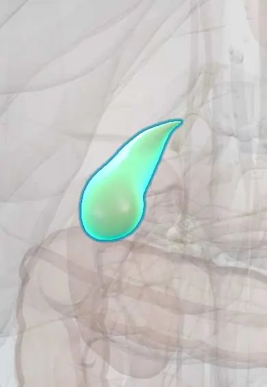
Gallbladder !
The gallbladder is a small pear-shaped sac under the liver, in the upper right belly, that stores bile; gallstones form in it.
Opens in the Durgical app, no sign-up needed.
What it is
A small pear-shaped sac under the liver that stores and concentrates bile.
What it does
Squeezes bile into the duodenum after fatty meals to help digest fat.
Key facts
- Where
- Gallbladder fossa on visceral liver surface (between segments IV and V); fundus at tip of right 9th costal cartilage, midclavicular line
- Blood supply
- Cystic artery (usually from right hepatic artery); cystic veins drain into liver bed and portal branches
- Nerve
- Hepatic plexus (celiac sympathetic, vagal); right phrenic nerve to its peritoneum (pain referred to right shoulder)
- Function
- Stores and concentrates bile (about 30–50 ml); contracts under CCK after fatty meals
Relations
- Above
- Gallbladder fossa on the visceral surface of the liver, between liver segment IV (IVb) and liver segment V, attached by connective tissue without peritoneum on that side
- In front
- The fundus projects beyond the lower edge of the liver against the anterior abdominal wall
- Below
- First and second parts of the duodenum and the transverse colon, which explains cholecystoduodenal and cholecystocolic fistulas
- Medial
- Neck near the porta hepatis and the hepatoduodenal ligament, where the cystic artery reaches it and the cystic duct leaves it
How to find it
The fundus lies where the lateral border of the right rectus abdominis meets the costal margin. A normal gallbladder cannot be felt; a large, smooth, non-tender one in a jaundiced person is the Courvoisier sign.
Examination
- Murphy sign: the examiner presses under the right costal margin at the gallbladder point while the person breathes in deeply; pain that cuts the breath short suggests acute cholecystitis
- Sonographic Murphy sign: most tenderness when the ultrasound probe presses right over the gallbladder seen on screen; supports acute cholecystitis
- Courvoisier sign: a palpable, non-tender gallbladder with jaundice points to a tumour below the cystic duct (often the pancreatic head) rather than stones, which leave a scarred, shrunken gallbladder
On imaging
Ultrasound first: stones are bright foci with posterior acoustic shadowing that move with position; acute cholecystitis adds wall thickening, fluid around the gallbladder and a sonographic Murphy sign. On hepatobiliary scintigraphy (HIDA), a gallbladder that never fills means a blocked cystic duct. CT misses many stones but shows complications.
Common problems
- Gallstones: very common (more in women, after pregnancy, with obesity, and in some Latin American populations); many cause no symptoms.
- Biliary colic: pain under the right ribs after meals when a stone blocks the outlet.
- Cholecystitis: an inflamed, infected gallbladder with fever.
How it is diagnosed
Ultrasound; blood tests.
Treatment
- Removal of the gallbladder by laparoscopy (cholecystectomy) for symptomatic stones.
Clinical notes
- Critical view of safety in laparoscopic cholecystectomy: the hepatocystic (Calot) triangle cleared of fat and fibrous tissue, the lower gallbladder lifted off the liver bed, and only two structures (cystic duct and cystic artery) entering it before anything is clipped
- Bile duct injury, usually from taking the common bile duct for the cystic duct, is the feared complication; inflammation and variant anatomy make it more likely
- Gallstone ileus: a large stone passes through a cholecystoduodenal fistula and blocks the terminal ileum; air in the bile ducts, small-bowel obstruction and an ectopic stone form Rigler triad
Anatomical variants
- Phrygian cap: a folded fundus, the most common shape variant; harmless, but it can mimic a septum on ultrasound
- Rare: double gallbladder, agenesis, an intrahepatic gallbladder, or a 'floating' gallbladder on a long mesentery that can twist
Development
Grows from the caudal part of the hepatic diverticulum of the foregut; its lumen is at first filled with cells and later hollows out, which helps explain septa and duplications.
Memory aid
Classic gallstone risk factors: female sex, pregnancies, age around 40 or more and obesity (the old English «4 Fs»), plus ancestry, rapid weight loss and family history
Related conditions
- Acute cholecystitis
- Gallstones (cholelithiasis)
- Gallbladder cancer
- Liver fluke infections (fascioliasis, clonorchiasis, opisthorchiasis)
- Intrahepatic cholestasis of pregnancy
Test yourself
Think of the answer, then tap the question to see it.
Where is the fundus of the gallbladder on the surface?
Where the lateral border of the right rectus abdominis meets the costal margin, at the tip of the right 9th costal cartilage.
What is Mirizzi syndrome?
A stone impacted in Hartmann pouch at the gallbladder neck presses on the common hepatic duct and causes jaundice.
What is the Courvoisier sign?
A large, smooth, non-tender gallbladder that can be felt in a jaundiced person; a normal gallbladder cannot be felt.
What is the critical view of safety in laparoscopic cholecystectomy?
Calot triangle cleared, lower gallbladder lifted off the liver bed, and only two structures (cystic duct and cystic artery) entering it before clipping.
Educational information, not medical advice. For any symptom, see a health professional.
Its names
- Official Latin (TA2)
- vesica biliaris TA2 3081
- Official Spanish (DeCS)
- Vesícula biliar DeCS D005704
- English (TA2)
- Gallbladder
Related
Sources: Terminologia Anatomica (TA2); DeCS; Anatomy, Abdomen and Pelvis: Gallbladder.
Spotted a mistake?We are always improving for the community. Tell us what is wrong and our team will review it.Report a mistake
Representative model, for educational purposes only.