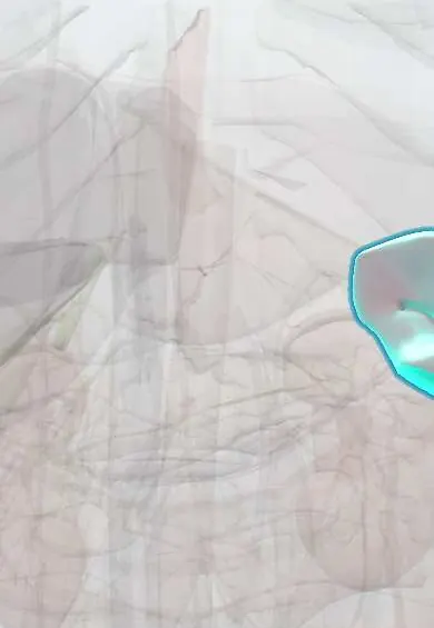
Spleen !
The spleen is a fist-sized organ in the upper left belly, under the lower ribs; it filters the blood and fights infection, and it bleeds easily when injured.
Opens in the Durgical app, no sign-up needed.
What it is
A fist-sized organ under the left ribs, behind the stomach.
What it does
Filters the blood, removes old red cells and helps fight some infections.
Key facts
- Where
- Left hypochondrium behind ribs 9–11, long axis along 10th rib; intraperitoneal, held by gastrosplenic and splenorenal ligaments
- Blood supply
- Splenic artery (celiac trunk); splenic vein to portal vein
- Nerve
- Splenic plexus from celiac plexus (mainly sympathetic)
- Function
- Filters blood and removes old red cells (red pulp); immune responses to blood-borne antigens (white pulp); platelet reservoir
Relations
- In front
- Stomach, joined by the gastrosplenic ligament, which carries the short gastric and left gastroepiploic vessels
- Behind
- Diaphragm, separating it from the left pleura, lung base and ribs; the left kidney and left adrenal gland
- Below
- Left (splenic) flexure of the colon and the phrenicocolic ligament, on which its lower pole rests
- Medial
- Tail of the pancreas at the hilum, in the splenorenal ligament with the splenic artery and splenic vein
How to find it
With the person supine, the examiner's left hand supports the lower left ribs from behind while the right hand starts low in the right iliac fossa and moves up toward the left costal margin as the person breathes in. Turning the person onto the right side helps bring a slightly enlarged spleen forward.
Examination
- Castell sign: the lowest left intercostal space in the anterior axillary line turns dull on percussion during full inspiration; suggests splenomegaly
- Traube space percussion: dullness over this left lower chest area, normally resonant from the stomach, suggests splenomegaly (or a left pleural effusion)
On imaging
Ultrasound measures length (splenomegaly is often taken as over about 12–13 cm; texts vary) and looks for free fluid in the left upper quadrant on FAST. Contrast CT grades splenic injury (AAST scale) and shows active bleeding as a contrast blush.
Common problems
- Rupture after an injury to the left ribs: internal bleeding.
- Enlarged spleen in infections, blood diseases or portal hypertension.
How it is diagnosed
Ultrasound or CT.
Treatment
- Observation, embolization or removal (splenectomy); vaccines afterwards because some infections become more dangerous.
Clinical notes
- Stable patients with splenic injury are often managed without surgery (observation or angioembolization) to keep its immune function
- Big spleens in tropical Latin America: malaria, visceral leishmaniasis and schistosomiasis (via portal hypertension), besides blood cancers and cirrhosis
- Splenic artery aneurysm is the most common visceral artery aneurysm; it is more frequent in women and can rupture in pregnancy
- Splenectomy can injure the tail of the pancreas at the hilum (pancreatic fistula) or the greater curvature of the stomach
Anatomical variants
- Accessory spleens (splenules) are common, usually near the hilum or in the gastrosplenic ligament; one left behind can cause relapse after splenectomy for immune thrombocytopenia
- Wandering spleen: lax or absent ligaments let it move and twist on its vascular pedicle
- Asplenia or polysplenia occur with heterotaxy syndromes
Development
Develops from mesenchyme in the dorsal mesogastrium, which is why it lies in the left upper quadrant held by the gastrosplenic and splenorenal ligaments (remnants of that mesentery); it is not a gut derivative, though the celiac trunk supplies it.
Memory aid
Odd numbers 1, 3, 5, 7, 9, 11: about 1 × 3 × 5 inches, about 7 ounces, lying between the 9th and 11th ribs.
Related conditions
- Splenic injury
- Pyruvate kinase deficiency
- Glucose-6-phosphate dehydrogenase (G6PD) deficiency
- Gaucher disease
- Niemann–Pick disease
- Hurler syndrome (mucopolysaccharidosis type I)
- Hunter syndrome (mucopolysaccharidosis type II)
- Sickle cell disease
- Thalassemias (α and β)
- Dengue
- Malaria
- Leishmaniasis
- Acute leukaemia (AML and ALL)
- Immune thrombocytopenia (ITP)
- Hodgkin lymphoma
- Non-Hodgkin lymphoma
- Chronic myeloid leukaemia
- Chronic lymphocytic leukaemia
- Autoimmune haemolytic anaemia
- Hereditary spherocytosis
- Polycythaemia vera
- Essential thrombocythaemia and primary myelofibrosis
- Asplenia and hyposplenism
- Mastocytosis (cutaneous and systemic)
- Infectious mononucleosis
- Histoplasmosis
- Typhoid (enteric) fever
- Brucellosis
- Viral haemorrhagic fevers (Ebola and others)
- Melioidosis
- Relapsing fever (tick-borne and louse-borne borreliosis)
- Babesiosis
- Wiskott–Aldrich syndrome
- Common variable immunodeficiency (CVID)
- Chédiak–Higashi syndrome
- Blunt abdominal trauma
Test yourself
Think of the answer, then tap the question to see it.
Behind which ribs does the spleen lie, and what holds it in place?
Ribs 9–11 in the left hypochondrium, its long axis along the 10th rib; it is intraperitoneal, held by the gastrosplenic and splenorenal ligaments.
What is Kehr sign in splenic rupture?
Pain at the tip of the left shoulder, from blood irritating the diaphragm, which is supplied by the phrenic nerve (C3–C5).
In which direction does an enlarged spleen grow, and what feature of its edge helps identify it?
Down and across toward the right iliac fossa, with a notched front edge; a normal spleen is not felt below the left costal margin.
Which infections are a lifelong risk after splenectomy?
Overwhelming infection by encapsulated bacteria: pneumococcus, meningococcus and Haemophilus influenzae type b; vaccination against them is part of care.
Which organ is at risk at the splenic hilum during splenectomy?
The tail of the pancreas, which lies at the hilum in the splenorenal ligament with the splenic vessels; injury causes a pancreatic fistula.
Educational information, not medical advice. For any symptom, see a health professional.
Its names
- Official Latin (TA2)
- splen TA2 5159
- Official Spanish (DeCS)
- Bazo DeCS D013154
- English (TA2)
- Spleen
Related
Sources: Terminologia Anatomica (TA2); DeCS; Anatomy, Abdomen and Pelvis, Spleen.
Spotted a mistake?We are always improving for the community. Tell us what is wrong and our team will review it.Report a mistake
Representative model, for educational purposes only.