Conditions · Metabolism: purines and pyrimidines
Hereditary xanthinuria !
An inherited disease in which xanthine oxidase is missing: uric acid almost disappears and xanthine forms kidney stones.
Updated 2026-11-01
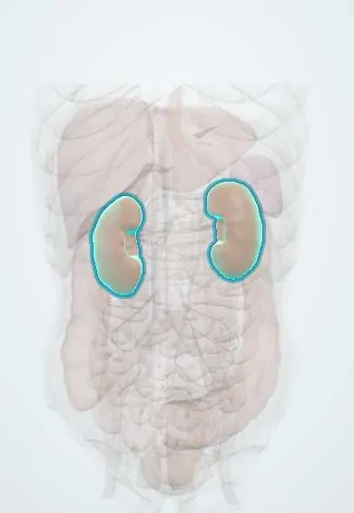
Structures involved
Tap one to see what it is and open it in 3D.
In biochemistry
What it is
Autosomal recessive deficiency of xanthine oxidoreductase (type I) or of it together with aldehyde oxidase (type II), with very low uric acid and xanthine build-up.
Who gets it
Estimated at about 1 in 69,000 people, although many cases go undiagnosed because they cause no symptoms; it seems more common in people of Mediterranean or Middle Eastern origin.
Causes
- Type I: XDH gene variants
- Type II: MOCOS gene variants (molybdenum cofactor sulfuration), which switch off xanthine oxidase and aldehyde oxidase
Risk factors
- Carrier parents or consanguinity
- Dehydration, which favours xanthine precipitation
Pathophysiology
Without xanthine oxidase, purine breakdown stops at hypoxanthine and xanthine. Hypoxanthine is partly recycled by HGPRT, but poorly soluble xanthine is excreted in urine and can precipitate as stones or deposit in kidneys and muscles. Blood and urine uric acid become almost undetectable.
Classification
- Type I (XDH)
- Type II (MOCOS)
- It must be told apart from molybdenum cofactor deficiency, which is far more severe, with neurological damage
Symptoms and signs
- Many people have no symptoms
- Renal colic and blood in the urine from xanthine stones
- Repeated urinary tract infections
- Muscle or joint pain from xanthine deposits
Diagnosis
- Clinical
- Radiolucent stones with very low uric acid in blood and urine.
- Tests
- Almost undetectable serum and urinary uric acid; high urinary xanthine and hypoxanthine; stone analysis (xanthine); allopurinol test to distinguish type I from type II; XDH and MOCOS genetic testing.
- Imaging
- Xanthine stones are not seen on plain X-ray; ultrasound and CT show them.
Treatment
- Lifestyle
- High fluid intake and a low-purine diet
- Procedure
- Stone removal by lithotripsy, ureteroscopy or percutaneous nephrolithotomy when they obstruct
- Follow-up
- Regular monitoring of kidney function and screening for new stones
Complications
- Urinary obstruction and infection
- Chronic kidney disease
- Myopathy from xanthine deposits
Prognosis
Good for most; the main risk is kidney damage from repeated stones.
Test yourself
Think of the answer, then tap the question to see it.
Which laboratory value points to xanthinuria?
Almost undetectable uric acid in blood and urine, with high urinary xanthine.
How do xanthine stones look on a plain X-ray?
They are not seen: they are radiolucent; ultrasound or CT detects them.
What distinguishes type II xanthinuria from type I?
In type II aldehyde oxidase is also missing (MOCOS defect), so allopurinol is not converted to oxypurinol.
Related conditions
Educational information, not medical advice. For any symptom, see a health professional.
Sources: Hereditary xanthinuria: MedlinePlus Genetics (MedlinePlus, US National Library of Medicine, 2026); XDH gene: MedlinePlus Genetics (MedlinePlus, US National Library of Medicine, 2026); XDH: Xanthine dehydrogenase/oxidase (human) (UniProt, 2026).
Spotted a mistake?We are always improving for the community. Tell us what is wrong and our team will review it.Report a mistake
Representative model, for educational purposes only.