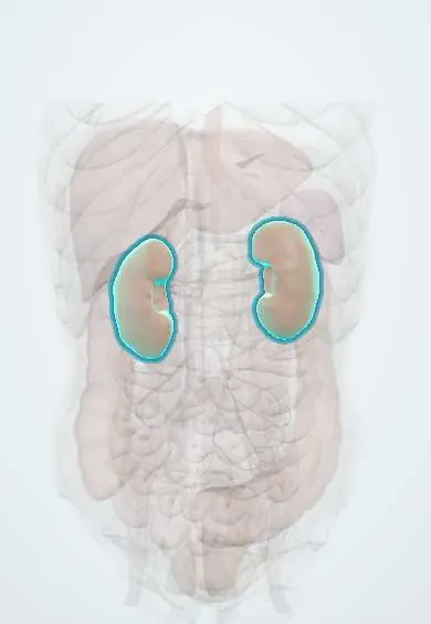
Kidney stones (urolithiasis) !
Kidney stones (urolithiasis) are hard crystals that form in the kidney; when one moves into the ureter it causes sudden, severe flank pain (renal colic).
Updated 2026-10-29
Structures involved
Tap one to see what it is and open it in 3D.
- Kidneys
- Renal pelvis
- Ureters
- Urinary bladder
- Common iliac arteries
- Psoas major
- Genitofemoral nerve
- Iliohypogastric and ilioinguinal nerves
In biochemistry
- Urinalysis
- Hypoxanthine-guanine phosphoribosyltransferase (HGPRT)
- Adenine phosphoribosyltransferase (APRT)
- Xanthine oxidase
- Uric acid
- Vitamin C (ascorbic acid)
- Calcium and phosphate
Microorganisms that cause it
In histology
In embryology
In the clinical exam
- Abdominal regions and quadrants: what lies under each
- Costovertebral angle tenderness (fist percussion)
- Urinary symptoms: dysuria, frequency, oliguria and hematuria
- Kidney palpation: bimanual palpation and ballottement
In physiology
- Patterns of malabsorption
- Glomerular filtration: the barrier and its determinants
- Renal handling of calcium, phosphate and magnesium
What it is
Formation of crystalline stones in the kidney (nephrolithiasis) that may move into the ureter, causing acute renal colic, blood in the urine and obstruction.
Who gets it
Common in adults of working age and frequently recurrent. Hot climates and dehydration increase risk, which matters in the warm lowland areas of Colombia. Historically more common in men, though the gap has narrowed.
Causes
- Calcium oxalate (most common) and calcium phosphate stones
- Uric acid stones (gout, metabolic syndrome, acidic urine)
- Struvite (infection) stones from urease-producing bacteria such as Proteus, often staghorn
- Cystine stones (cystinuria, inherited)
- Drug-related stones
Risk factors
- Low fluid intake, hot climate and dehydration
- Diet high in sodium and animal protein; high oxalate intake
- Obesity, diabetes, metabolic syndrome and gout
- Primary hyperparathyroidism, renal tubular acidosis, bowel disease or surgery (enteric hyperoxaluria)
- Previous stones or family history
- Anatomical problems: horseshoe kidney, ureteropelvic junction obstruction
Pathophysiology
Urine supersaturated with stone-forming salts, plus low inhibitors such as citrate, lets crystals nucleate and grow, often on papillary plaques. A stone that enters the ureter tends to lodge at narrow points: the ureteropelvic junction, the crossing of the iliac vessels and the ureterovesical junction. Obstruction raises pressure in the collecting system and stretches the capsule, causing colic.
Classification
- By composition: calcium oxalate, calcium phosphate, uric acid, struvite, cystine
- By location: renal (calyceal, pelvic, staghorn), ureteral (proximal, mid, distal), bladder
- By imaging: radio-opaque vs radiolucent (uric acid) on plain film
- Obstructing with infection (urological emergency) vs uncomplicated
Symptoms and signs
- Sudden severe colicky flank pain radiating to the groin, testicle or labium
- Restlessness: the patient cannot find a comfortable position
- Nausea and vomiting
- Visible or microscopic haematuria
- Urinary frequency, urgency or pain on passing urine when the stone is near the bladder
- Some stones are found by chance without symptoms
Diagnosis
- Clinical
- Typical colicky loin-to-groin pain in a restless patient, with costovertebral angle tenderness and a soft abdomen. Check temperature, blood pressure and pregnancy status.
- Tests
- Urinalysis (blood, infection signs, pH), urine culture if infection is suspected, blood count, creatinine, electrolytes, calcium and uric acid; pregnancy test. Stone analysis and, in recurrent formers, 24-hour urine studies.
- Imaging
- Non-contrast low-dose CT of abdomen and pelvis is the reference test: it shows stone size, site and hydronephrosis. Ultrasound first in pregnancy and children and to show hydronephrosis; plain film (KUB) to follow radio-opaque stones.
Differential diagnosis
- Acute pyelonephritis
- Acute appendicitis or diverticulitis
- Biliary colic
- Ectopic pregnancy, ovarian torsion or cyst
- Abdominal aortic aneurysm
- Testicular torsion, musculoskeletal back pain
Goal of treatment
Control pain, recognise and urgently drain an infected or obstructed kidney, help the stone pass or remove it, and prevent recurrence.
Treatment
- Acute
- Analgesia: NSAIDs are first-line when kidney function allows; opioids and antiemetics if needed.
- Medical
- Medical expulsive therapy with an alpha-blocker for distal ureteral stones of a size likely to pass, with observation; small stones usually pass spontaneously and larger ones are less likely to.
- Acute
- An obstructed, infected kidney is an emergency: urgent decompression (ureteral stent or percutaneous nephrostomy) plus antibiotics, with definitive stone treatment later.
- Procedure
- Stone removal when it will not pass or causes problems: shock wave lithotripsy, ureteroscopy with laser lithotripsy, or percutaneous nephrolithotomy for large or staghorn stones.
- Lifestyle
- Prevention: drinking enough to produce plenty of urine, less sodium and animal protein, normal dietary calcium.
- Medical
- Drugs by stone type in recurrent formers: thiazide diuretics (hypercalciuria), citrate (hypocitraturia, uric acid stones), xanthine oxidase inhibitors (hyperuricosuria); treatment of hyperparathyroidism.
Drugs for this condition
Complications
- Obstructive pyelonephritis, pyonephrosis and urosepsis
- Hydronephrosis and loss of kidney function
- Acute kidney injury (single kidney or bilateral obstruction)
- Ureteral stricture
- Recurrence
- Chronic kidney disease with repeated or staghorn stones
Prognosis
Most small ureteral stones pass with supportive care, and intervention clears most of the rest. Recurrence is common without prevention, so metabolic assessment is advised for recurrent formers.
Prevention
- Adequate daily fluid intake, especially in hot climates
- Reduced salt and animal protein; normal calcium intake
- Weight control and management of diabetes and gout
- Targeted drug therapy after stone analysis
Test yourself
Think of the answer, then tap the question to see it.
What is the imaging test of choice for suspected renal colic in non-pregnant adults?
Non-contrast low-dose CT of the abdomen and pelvis.
At which three sites do ureteral stones most often lodge?
The ureteropelvic junction, where the ureter crosses the iliac vessels, and the ureterovesical junction.
Which stone is radiolucent on plain X-ray?
Uric acid stones (pure); they are still visible on CT.
What is the management of an obstructing stone with fever?
Urgent decompression with a ureteral stent or percutaneous nephrostomy plus antibiotics; definitive stone treatment is delayed.
Which organism is classically linked to struvite (staghorn) stones?
Urease-producing bacteria, especially Proteus, which alkalinise the urine.
Related conditions
- Acute pyelonephritis
- Acute appendicitis
- Abdominal aortic aneurysm
- Testicular torsion
- Ectopic pregnancy
- Diverticular disease and diverticulitis
- Crohn disease
- Chronic kidney disease
Educational information, not medical advice. For any symptom, see a health professional.
Sources: Renal Calculi, Nephrolithiasis (StatPearls (NCBI Bookshelf), 2024).
Spotted a mistake?We are always improving for the community. Tell us what is wrong and our team will review it.Report a mistake
Representative model, for educational purposes only.