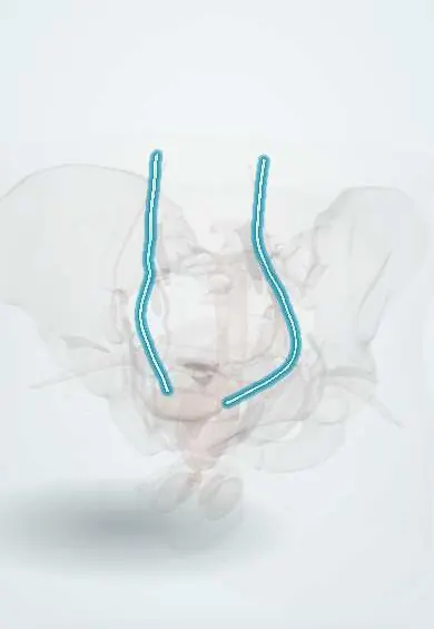
Ureters !
The ureters are two thin muscular tubes that carry urine from the kidneys to the bladder in waves; kidney stones can get stuck in them.
Official name: Ureter
Opens in the Durgical app, no sign-up needed.
What it is
The two tubes that carry urine from the kidneys to the bladder. In the pelvis each runs under the uterine artery, about 1.5-2 cm to the side of the cervix.
What it does
Move urine down by waves of contraction.
Key facts
- Where
- Retroperitoneal, from renal pelvis (about L2) down on psoas major, over the pelvic brim at the common iliac bifurcation, to the bladder; about 25 cm
- Blood supply
- Branches of renal, gonadal, aortic, common and internal iliac, and vesical arteries
- Nerve
- Renal, aortic and hypogastric plexuses; pain fibres T11–L2 (loin-to-groin pain)
- Function
- Carry urine from kidney to bladder by peristalsis; narrowest at pelviureteric junction, pelvic brim and entry to bladder
Relations
- Course
- From the renal pelvis it descends retroperitoneally on psoas major, enters the pelvis over the common iliac bifurcation, runs down the side wall in front of the internal iliac artery, then turns forward and medially to the bladder base
- In front
- Abdomen: the gonadal vessels cross it; the duodenum (right) or the sigmoid mesocolon (left). Pelvis: the uterine artery (female) or the ductus deferens (male) arches over it
- Behind
- Psoas major and the genitofemoral nerve, then the bifurcation of the common iliac artery at the pelvic brim
- Medial
- Inferior vena cava (right ureter) and abdominal aorta (left ureter); in the pelvis, the peritoneum of the side wall
How to find it
Not palpable. At surgery it is found at the pelvic brim crossing the iliac bifurcation, under the peritoneum of the side wall, and recognised by its peristalsis when touched; in the female it is traced down to the uterine artery before pelvic vessels are clamped.
On imaging
Non-contrast CT is the standard test for stones; CT urography shows the whole course and any leak. On a plain film its line runs near the tips of the lumbar transverse processes, over the sacroiliac joint and toward the ischial spine; pelvic phleboliths can mimic stones.
Common problems
- Can be injured in a hysterectomy or other pelvic surgery ('water under the bridge': the ureter runs under the uterine artery).
- Kidney stones can get stuck in them.
How it is diagnosed
CT urography; checks during surgery when injury is suspected.
Treatment
- Surgeons identify and protect them; an injury is repaired, sometimes with a temporary stent.
Clinical notes
- Its blood comes from the medial side in the abdomen and the lateral side in the pelvis, so surgeons keep its sheath (adventitia) intact when mobilising it
- Female: it forms the posterior border of the ovarian fossa, so ovarian masses and side-wall endometriosis can involve it
- An unrecognised surgical injury can present days later with flank pain, fever or urine leaking from the vagina (ureterovaginal fistula)
- Large pelvic tumours, cervical cancer or a pregnant uterus can compress it and cause hydronephrosis, more often on the right in pregnancy
Anatomical variants
- Duplex ureter, partial (bifid) or complete, one of the commonest urinary tract anomalies
- Congenital pelviureteric junction obstruction, a common cause of hydronephrosis found before birth
- Retrocaval ureter (right side, passing behind the inferior vena cava), rare
Development
Grows from the ureteric bud, an outgrowth of the mesonephric duct that also induces the kidney; early splitting of the bud gives a bifid or duplex ureter.
Related conditions
- Kidney stones (urolithiasis)
- Acute pyelonephritis
- Acute kidney injury
- Hydronephrosis and obstructive uropathy
- Vesicoureteral reflux
- Congenital kidney anomalies (horseshoe kidney, renal agenesis)
Test yourself
Think of the answer, then tap the question to see it.
Where do ureteric stones most often lodge?
At the ureterovesical junction, as the ureter passes through the bladder wall, one of its narrowings.
Which structure crosses over the pelvic ureter in the female and in the male?
The uterine artery in the female and the ductus deferens in the male («water under the bridge»).
What are the classic surgical injury sites of the ureter?
The pelvic brim beside the suspensory ligament of the ovary, the uterine artery crossing and near the vaginal vault.
Why do surgeons keep the ureteric adventitia intact when mobilising the ureter?
Its blood comes from the medial side in the abdomen and the lateral side in the pelvis, running in the sheath.
How may an unrecognised surgical ureteric injury present?
Days later with flank pain, fever or urine leaking from the vagina (ureterovaginal fistula).
Educational information, not medical advice. For any symptom, see a health professional.
Its names
- Official Latin (TA2)
- ureter TA2 3394
- Official Spanish (DeCS)
- Uréter DeCS D014513
- English (TA2)
- Ureter
Related
Sources: Terminologia Anatomica (TA2); DeCS; Anatomy, Abdomen and Pelvis Ureter.
Spotted a mistake?We are always improving for the community. Tell us what is wrong and our team will review it.Report a mistake
Representative model, for educational purposes only.