Conditions · Ophthalmology and ENT
Optic neuritis !
Optic neuritis is inflammation of the optic nerve, causing painful vision loss in one eye over days. It is often the first sign of multiple sclerosis.
Updated 2026-11-01
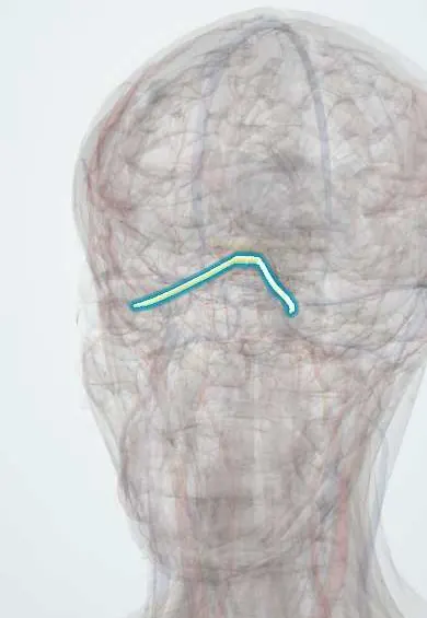
Structures involved
Tap one to see what it is and open it in 3D.
Why it happens
What it is
Inflammatory demyelination of the optic nerve that causes subacute vision loss in one eye, usually with pain on eye movement. The typical form is closely linked to multiple sclerosis.
Who gets it
Most common in women aged 20–45. In Latin America, including Colombia, a higher proportion of cases than in Europe are linked to neuromyelitis optica spectrum disorder (aquaporin-4 antibodies), which matters because its prognosis and treatment differ.
Causes
- Idiopathic or multiple sclerosis-related demyelination (typical optic neuritis)
- Neuromyelitis optica spectrum disorder (aquaporin-4 IgG) and MOG antibody-associated disease
- Infections: syphilis, tuberculosis, viral infections (including zoster), Lyme disease
- Systemic inflammatory disease: sarcoidosis, systemic lupus erythematosus
Risk factors
- Female sex and young adult age
- Multiple sclerosis or previous demyelinating episodes
- Recent viral infection (especially in children)
Pathophysiology
Immune-mediated inflammation strips the myelin from optic nerve axons, slowing or blocking conduction. Swelling of the nerve within its sheath stretches the dura at the orbital apex, which explains pain on eye movement. Remyelination allows recovery over weeks, but some axonal loss leaves optic disc pallor.
Classification
- Retrobulbar (normal disc, about two thirds of adults) vs papillitis (swollen disc)
- Typical (multiple sclerosis-associated) vs atypical (neuromyelitis optica, MOG, infectious, inflammatory)
Symptoms and signs
- Loss of vision in one eye over hours to days
- Pain behind the eye, worse on eye movement
- Reduced colour vision (red desaturation) and contrast
- Central or paracentral blind spot (scotoma)
- Vision worse with heat or exercise (Uhthoff phenomenon)
Diagnosis
- Clinical
- Reduced visual acuity and colour vision, a relative afferent pupillary defect (Marcus Gunn pupil) in the affected eye, central visual field defect, and a normal or swollen optic disc on fundoscopy.
- Tests
- Visual field testing; aquaporin-4 and MOG antibodies in atypical cases; syphilis serology and other infectious or inflammatory tests when suggested; CSF analysis in selected patients.
- Imaging
- Brain and orbit MRI with contrast shows optic nerve enhancement and looks for white-matter lesions, which predict the risk of multiple sclerosis. Optical coherence tomography follows nerve fibre loss.
Differential diagnosis
- Non-arteritic or arteritic (giant cell arteritis) ischaemic optic neuropathy
- Compressive optic neuropathy (tumour, aneurysm)
- Papilloedema
- Leber hereditary optic neuropathy
- Toxic or nutritional optic neuropathy
Goal of treatment
Speed recovery of vision, identify atypical causes that need specific treatment, and assess and reduce the risk of multiple sclerosis.
Treatment
- Medical
- High-dose intravenous corticosteroids speed visual recovery but do not change final vision; oral corticosteroids alone at standard doses were linked to more recurrences in trials.
- Medical
- Plasma exchange for severe attacks not responding to corticosteroids, especially in neuromyelitis optica.
- Medical
- Disease-modifying therapy when MRI or clinical findings indicate multiple sclerosis; long-term immunosuppression for neuromyelitis optica and MOG disease.
- Follow-up
- Follow-up of visual acuity, fields and colour vision, and neurology review for new symptoms.
Complications
- Residual reduced colour vision, contrast or acuity; optic disc pallor
- Recurrence in the same or the other eye
- Development of multiple sclerosis
- Corticosteroid side effects
Prognosis
Typical optic neuritis has a good outlook: most patients recover most of their vision within weeks to a few months, even without treatment. The risk of multiple sclerosis depends mainly on MRI brain lesions. Neuromyelitis optica-related optic neuritis often leaves severe deficits.
Test yourself
Think of the answer, then tap the question to see it.
What is the classic triad of typical optic neuritis?
Subacute unilateral vision loss, pain on eye movement and reduced colour vision, with a relative afferent pupillary defect.
Which test best predicts the risk of multiple sclerosis after optic neuritis?
Brain MRI: white-matter demyelinating lesions mean a much higher risk.
What did high-dose intravenous corticosteroids achieve in the Optic Neuritis Treatment Trial?
Faster recovery of vision without changing the final visual outcome.
Which antibody points to neuromyelitis optica in a patient with severe optic neuritis?
Aquaporin-4 IgG (anti-AQP4).
Related conditions
- Multiple sclerosis
- Neuromyelitis optica spectrum disorder
- Giant cell arteritis
- Idiopathic intracranial hypertension
- Sarcoidosis
- Systemic lupus erythematosus
- Syphilis
Educational information, not medical advice. For any symptom, see a health professional.
Sources: Optic neuritis: MedlinePlus Medical Encyclopedia (MedlinePlus (U.S. National Library of Medicine), 2024).
Spotted a mistake?We are always improving for the community. Tell us what is wrong and our team will review it.Report a mistake
Representative model, for educational purposes only.