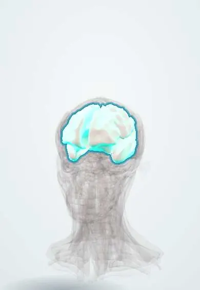
Multiple sclerosis !
Multiple sclerosis is an immune disease that damages the nerve coating in the brain, optic nerves and spinal cord, causing episodes of nerve symptoms.
Updated 2026-10-29
Structures involved
Tap one to see what it is and open it in 3D.
- White matter
- Optic nerve
- Spinal cord
- Corpus callosum
- Lateral ventricles
- Pons
- Cerebellum
- Cerebellar peduncles
- Medulla oblongata
- Trigeminal nerve (V)
In biochemistry
In immunology
- Type IV hypersensitivity (delayed, T cells)
- HLA associations with disease
- Molecular mimicry and post-infectious autoimmunity
- Lymphocyte recirculation and homing
- Type I interferons (α, β)
- Adhesion molecules: selectins and integrins
In histology
- Glial cells: astrocytes, oligodendrocytes, microglia, Schwann cells
- Gray and white matter, cerebellar cortex, meninges
- Blood–brain barrier, choroid plexus and cerebrospinal fluid
In the clinical exam
- Cranial nerve examination
- Babinski sign
- Hoffmann sign and clonus
- Cerebellar tests: finger–nose, heel–shin and rapid alternating movements
- Pupils: light reflex and abnormal pupils (Marcus Gunn, Horner, Argyll Robertson)
- Upper vs lower motor neuron syndrome
- Spinal cord syndromes: complete, Brown-Séquard, central, anterior
- Cerebellar syndrome
In physiology
- Impulse conduction and myelin
- Somatosensory pathways: dorsal column and anterolateral
- Upper versus lower motor neuron lesions
- Functions of the cerebellum
- The visual pathway, pupillary reflexes and eye movements
Why it happens
What it is
A chronic immune-mediated disease of the central nervous system in which inflammation destroys myelin and axons in the brain, optic nerves and spinal cord. It causes episodes of neurological symptoms separated in time and place, with possible progression.
Who gets it
Usually starts between about 20 and 40 years of age and is two to three times more common in women. It is less frequent in Latin America than in northern Europe, but it is a leading cause of non-traumatic disability in young adults.
Causes
- Autoimmune attack on central nervous system myelin, triggered in genetically susceptible people
- Epstein-Barr virus infection strongly associated
- Genetic susceptibility (HLA-DRB1 and other variants)
Risk factors
- Female sex
- Young adulthood
- Prior Epstein-Barr virus infection, particularly infectious mononucleosis
- Low vitamin D or little sun exposure
- Smoking
- Obesity in adolescence
- Family history
Pathophysiology
Autoreactive T and B lymphocytes cross the blood-brain barrier and cause inflammation, demyelination and axonal loss. Plaques favour periventricular white matter, the corpus callosum, juxtacortical areas, brainstem, cerebellum, optic nerves and spinal cord. Remyelination is incomplete; accumulated axonal loss drives permanent disability.
Classification
- Clinically isolated syndrome
- Relapsing-remitting (most common at onset)
- Secondary progressive
- Primary progressive
- Each described as active or not, and progressing or not
Symptoms and signs
- Optic neuritis: painful loss of vision in one eye, worse with eye movement
- Numbness, tingling or weakness of limbs
- Lhermitte sign: electric sensation down the spine on neck flexion
- Double vision (internuclear ophthalmoplegia)
- Ataxia, tremor, vertigo
- Bladder and bowel dysfunction, fatigue, heat sensitivity (Uhthoff phenomenon), cognitive and mood changes
Diagnosis
- Clinical
- Demonstration of dissemination in space and time of central nervous system lesions using the McDonald criteria, combining clinical attacks, MRI and cerebrospinal fluid, after excluding other causes.
- Tests
- Cerebrospinal fluid: oligoclonal bands specific to CSF support the diagnosis. Visual evoked potentials. Blood tests to exclude mimics (e.g. aquaporin-4 and MOG antibodies, vitamin B12, HIV, syphilis).
- Imaging
- MRI of brain and spinal cord with gadolinium: T2 lesions in typical locations (periventricular, juxtacortical, infratentorial, spinal cord); enhancing and non-enhancing lesions together show dissemination in time.
Differential diagnosis
- Neuromyelitis optica spectrum disorder and MOG antibody disease
- Acute disseminated encephalomyelitis
- Small-vessel ischaemic disease
- Vitamin B12 deficiency
- Infections (HIV, syphilis, HTLV-1 myelopathy)
- Systemic lupus and other vasculitides
Goal of treatment
Shorten relapses, reduce new relapses and MRI activity, delay disability and manage symptoms.
Treatment
- Acute
- Relapses: high-dose corticosteroids for a short course; plasma exchange for severe relapses that do not respond.
- Medical
- Disease-modifying therapies started early: injectable immunomodulators, oral agents and monoclonal antibodies, chosen by activity, risks and access.
- Medical
- Symptomatic treatment of spasticity, neuropathic pain, bladder dysfunction, fatigue and depression.
- Rehabilitation
- Physiotherapy, occupational therapy and exercise to preserve mobility and function.
- Follow-up
- Regular clinical review and MRI to monitor activity and treatment safety.
Drugs for this condition
Complications
- Accumulated disability and loss of mobility
- Urinary tract infections and bladder dysfunction
- Depression and cognitive impairment
- Pressure ulcers and falls in advanced disease
- Infections or other adverse effects of immunotherapy
Prognosis
Highly variable. Many people keep good function for years, especially with early disease-modifying therapy. Male sex, older onset, motor or cerebellar onset, frequent early relapses and high MRI lesion load predict a worse course.
Test yourself
Think of the answer, then tap the question to see it.
Which principle underlies the diagnosis of multiple sclerosis?
Dissemination of central nervous system lesions in space and in time (McDonald criteria), after excluding other causes.
Which cerebrospinal fluid finding supports multiple sclerosis?
Oligoclonal bands present in CSF and absent in serum.
What is Lhermitte sign?
An electric shock-like sensation down the spine or limbs on flexing the neck, from cervical spinal cord lesions.
What is the classic presentation of optic neuritis?
Subacute painful loss of vision in one eye, worse on eye movement, with reduced colour vision.
Where do multiple sclerosis plaques typically appear on brain MRI?
Periventricular white matter (including the corpus callosum), juxtacortical areas, infratentorial regions and the spinal cord.
Related conditions
Educational information, not medical advice. For any symptom, see a health professional.
Sources: Multiple Sclerosis (StatPearls (NCBI Bookshelf), 2024).
Spotted a mistake?We are always improving for the community. Tell us what is wrong and our team will review it.Report a mistake
Representative model, for educational purposes only.