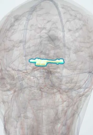
Nervous system · Head and neck
Optic chiasm !
The optic chiasm is where the two optic nerves meet and partly cross, just above the pituitary; a pituitary tumour here narrows side vision.
Opens in the Durgical app, no sign-up needed.
What it is
The X where the two optic nerves meet above the pituitary, then split into the optic tracts.
What it does
Fibres from the inner half of each retina cross here; a pituitary tumour pressing up causes tunnel vision on both sides.
Key facts
- Roots
- Optic nerves meet above the sella turcica
- Moves
- None
- Feeling from
- Nasal retina fibres cross (temporal visual fields): bitemporal hemianopia if compressed
Relations
- In front
- Optic nerves converging from the optic canals
- Below
- Diaphragma sellae and the pituitary gland in the sella turcica, with the sphenoid sinus below
- Lateral
- Supraclinoid internal carotid arteries; the cavernous sinuses lie lower down on each side of the sella
- Above
- Floor of the third ventricle (optic recess) and the lamina terminalis; above and in front, the anterior cerebral arteries and the anterior communicating artery
- Behind
- Pituitary stalk (infundibulum), tuber cinereum and mammillary bodies; the optic tracts diverge backward
How to find it
On coronal MRI it is the flat band above the pituitary gland and the diaphragma sellae, with the third ventricle above and behind and the internal carotid arteries at its sides. In a pterional approach it is seen between the two optic nerves once the sylvian fissure is opened.
Examination
- Confrontation fields, each eye tested separately: a temporal defect that stops at the vertical midline points to the chiasm
- Automated perimetry maps defects that respect the vertical meridian and is repeated to follow a tumour or its treatment
On imaging
MRI of the sella with contrast, in coronal and sagittal planes, shows the chiasm draped over or lifted by a pituitary adenoma; OCT can show thinning of the nasal retinal fibres.
Clinical notes
- Pituitary adenomas may also cause hormonal syndromes (high prolactin, acromegaly, Cushing disease) or loss of pituitary function
- Pituitary apoplexy: bleeding into an adenoma with sudden headache, loss of vision and double vision; an emergency
- Endoscopic surgery through the nose and sphenoid sinus reaches the pituitary from below, decompressing the chiasm
Anatomical variants
- Most chiasms lie directly above the diaphragma sellae; a prefixed chiasm (further forward) makes pituitary tumours hit the optic tracts, and a postfixed one makes them hit the optic nerves
Development
It forms where the two optic nerves meet and partly cross as retinal fibres grow back toward the diencephalon. In albinism more fibres than usual cross, which disturbs binocular vision; in septo-optic dysplasia the optic nerves and chiasm are small.
Related conditions
Test yourself
Think of the answer, then tap the question to see it.
Which visual field defect does compression of the optic chiasm from below by a pituitary macroadenoma cause?
Bitemporal hemianopia, starting in the upper temporal quadrants, because the crossing lower nasal fibres are hit first.
Which tumour presses on the optic chiasm from above, and which quadrants tend to go first?
Craniopharyngioma, often in children; the lower temporal quadrants tend to be lost first.
What does pressure on the side of the optic chiasm, for example from an internal carotid aneurysm, cause?
A nasal field defect in the eye on that side, because it affects the uncrossed temporal fibres.
What is pituitary apoplexy and why does it threaten the optic chiasm?
Bleeding into a pituitary adenoma: sudden headache, loss of vision and double vision; an emergency because the mass presses up on the chiasm.
Educational information, not medical advice. For any symptom, see a health professional.
Its names
- Official Latin (TA2)
- chiasma opticum TA2 5668
- Official Spanish (DeCS)
- Quiasma Óptico DeCS D009897
- English (TA2)
- Optic chiasm
- Also called
- optic chiasma
Related
Sources: Terminologia Anatomica (TA2); DeCS; Neuroanatomy, Optic Chiasm; Anatomy of the Human Body (20th ed.): Development of the Nervous System (Lea & Febiger (Gray's Anatomy, 1918), 1918) · adapted, Public domain.
Spotted a mistake?We are always improving for the community. Tell us what is wrong and our team will review it.Report a mistake
Representative model, for educational purposes only.