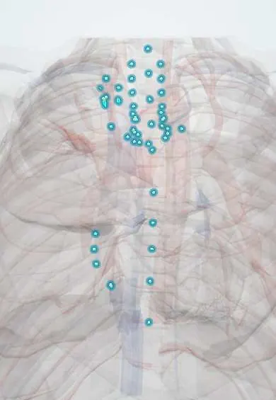
Sarcoidosis !
Sarcoidosis is an immune disease that forms small inflammatory clumps (granulomas), most often in the lungs and chest lymph nodes.
Updated 2026-11-01
Structures involved
Tap one to see what it is and open it in 3D.
In immunology
Why it happens
- Granulomatous inflammation: caseating vs non-caseating, the causes to know
- Pathologic calcification: dystrophic vs metastatic
- Calcium regulation: PTH, vitamin D and calcitonin
- Obstructive and restrictive patterns, and flow–volume loops
- Vitamin D (calcitriol)
What it is
A multisystem inflammatory disease of unknown cause defined by non-caseating granulomas. The lungs and the hilar and mediastinal lymph nodes are involved in most patients; skin, eyes, heart, nervous system, liver and kidneys can also be affected.
Who gets it
Usually begins between 20 and 50 years of age, with a second peak in women over 50. It is more frequent and more severe in people of African descent and in Northern Europeans. In Colombia and Latin America it is less often diagnosed than in Europe, partly because tuberculosis and endemic fungal infections cause similar granulomas and must be excluded first.
Causes
- Unknown cause; thought to be an exaggerated immune response to an unidentified antigen in a genetically susceptible person
- Suspected triggers include environmental dusts, metals and mycobacterial or other microbial antigens
- Genetic factors (HLA variants) and family history increase susceptibility
Risk factors
- Age 20–50 years
- African descent or Northern European ancestry
- Female sex
- Family history of sarcoidosis
- Occupational exposure to dusts, metals, mould or agricultural products
Pathophysiology
Antigen-presenting cells activate CD4+ T helper 1 lymphocytes, which release cytokines (interferon-gamma, tumour necrosis factor) and recruit macrophages that fuse into epithelioid and giant cells. These form tight, non-caseating granulomas along lymphatics, bronchovascular bundles and the pleura. Most granulomas resolve; persistent ones lead to fibrosis, mainly in the upper lobes. Activated macrophages convert vitamin D to its active form, which explains hypercalcaemia and hypercalciuria, and raise serum angiotensin-converting enzyme (ACE).
Classification
- Scadding chest X-ray stage 0: normal film
- Stage I: bilateral hilar lymphadenopathy only
- Stage II: hilar lymphadenopathy plus lung infiltrates
- Stage III: lung infiltrates without lymphadenopathy
- Stage IV: lung fibrosis (honeycombing, upper-lobe retraction)
Symptoms and signs
- Dry cough, shortness of breath and chest discomfort; many patients have no symptoms and are found on a chest X-ray
- Fatigue, fever, night sweats and weight loss
- Skin lesions: erythema nodosum on the shins or lupus pernio (violaceous plaques on the nose and cheeks)
- Red, painful eyes or blurred vision from uveitis
- Löfgren syndrome: fever, erythema nodosum, bilateral hilar adenopathy and ankle arthritis
- Palpitations, syncope or facial palsy when the heart or nerves are involved
Diagnosis
- Clinical
- Diagnosis rests on three steps: a compatible clinical and radiological picture, tissue showing non-caseating granulomas, and exclusion of other granulomatous diseases (tuberculosis, histoplasmosis, lymphoma, pneumoconiosis). Löfgren syndrome is typical enough that biopsy is often not needed.
- Tests
- Serum calcium, 24-hour urinary calcium, kidney and liver function; serum ACE is often raised but is neither sensitive nor specific. Tuberculin skin test or interferon-gamma release assay (often anergic). Spirometry and diffusing capacity (restrictive pattern, low DLCO). ECG in every patient. Biopsy by endobronchial ultrasound-guided needle aspiration of mediastinal nodes or transbronchial biopsy; bronchoalveolar lavage shows a high CD4/CD8 ratio. Mycobacterial and fungal stains and cultures of the tissue.
- Imaging
- Chest X-ray: bilateral symmetrical hilar lymphadenopathy is the classic sign. High-resolution CT: perilymphatic nodules along fissures and bronchovascular bundles, upper-lobe fibrosis. Ophthalmological slit-lamp examination; cardiac MRI or PET when the heart is suspected.
Differential diagnosis
- Pulmonary tuberculosis (caseating granulomas)
- Endemic fungal infections: histoplasmosis, paracoccidioidomycosis
- Lymphoma (Hodgkin and non-Hodgkin)
- Hypersensitivity pneumonitis and berylliosis or other pneumoconioses
- Metastatic cancer to the mediastinal nodes
Goal of treatment
Control inflammation only when it threatens organ function or causes significant symptoms, preserve lung, heart, eye and nerve function, and avoid unnecessary long-term immunosuppression.
Treatment
- Follow-up
- Many patients, especially stage I or Löfgren syndrome, improve without treatment and are followed with symptoms, chest X-ray, spirometry, ECG and calcium.
- Medical
- Systemic glucocorticoids are first line when treatment is needed (symptomatic or progressive lung disease, cardiac, eye, nervous system or hypercalcaemic involvement).
- Medical
- Steroid-sparing agents (methotrexate, azathioprine, mycophenolate) or anti-TNF biologic therapy in chronic or refractory disease; topical corticosteroid eye drops for anterior uveitis.
- Procedure
- Pacemaker or implantable cardioverter-defibrillator in cardiac sarcoidosis with heart block or ventricular arrhythmias; lung transplantation in end-stage fibrosis.
Complications
- Pulmonary fibrosis and pulmonary hypertension
- Heart block, ventricular arrhythmias and sudden death
- Uveitis, glaucoma, cataract and vision loss
- Hypercalcaemia, kidney stones and kidney failure
- Aspergilloma in fibrotic cavities
Prognosis
Most patients have spontaneous remission within two to three years, especially those with Löfgren syndrome or stage I disease. About a quarter develop chronic disease; advanced fibrosis, pulmonary hypertension and cardiac involvement are the main causes of death.
Prevention
- No known primary prevention
- Reduction of occupational dust and metal exposure
- Bone protection and infection screening during long glucocorticoid treatment
Test yourself
Think of the answer, then tap the question to see it.
What is the classic chest X-ray finding in sarcoidosis?
Bilateral symmetrical hilar lymphadenopathy, with or without lung infiltrates (Scadding stage I or II).
What histological finding defines sarcoidosis?
Non-caseating epithelioid granulomas, after tuberculosis, fungal infection and other causes of granulomas are excluded.
Why can patients with sarcoidosis develop hypercalcaemia?
Activated macrophages in granulomas convert vitamin D to calcitriol, which increases intestinal calcium absorption.
What is Löfgren syndrome?
An acute form of sarcoidosis with fever, erythema nodosum, bilateral hilar adenopathy and ankle arthritis; it usually has a good prognosis.
What is first-line treatment when sarcoidosis needs therapy?
Systemic glucocorticoids; steroid-sparing immunosuppressants or anti-TNF agents are added for chronic or refractory disease.
Related conditions
- Pulmonary tuberculosis
- Histoplasmosis
- Hodgkin lymphoma
- Non-Hodgkin lymphoma
- Interstitial lung disease
- Hypersensitivity pneumonitis
- Pneumoconioses (silicosis, asbestosis, coal workers' pneumoconiosis)
- Uveitis
- Erythema nodosum
- Atrioventricular (heart) block
- Bell palsy
- Kidney stones (urolithiasis)
- Pulmonary hypertension and cor pulmonale
Educational information, not medical advice. For any symptom, see a health professional.
Sources: Sarcoidosis (MedlinePlus Medical Encyclopedia (U.S. National Library of Medicine), 2024); What Is Sarcoidosis? (National Heart, Lung, and Blood Institute (NHLBI, NIH), 2022).
Spotted a mistake?We are always improving for the community. Tell us what is wrong and our team will review it.Report a mistake
Representative model, for educational purposes only.