Neuromyelitis optica spectrum disorder !
An autoimmune disease that attacks the optic nerves and spinal cord, causing attacks of vision loss and paralysis. Preventing relapses is key.
Updated 2026-11-01
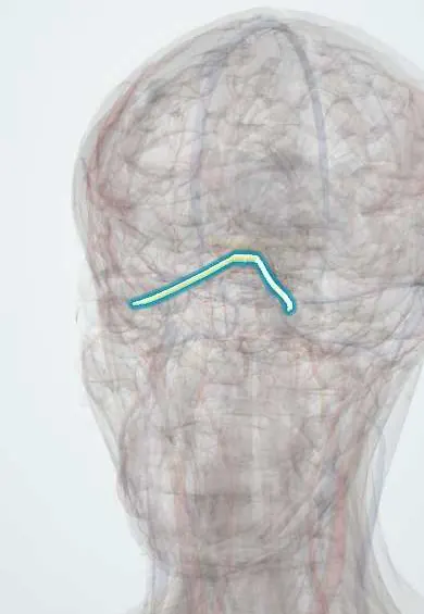
Structures involved
Tap one to see what it is and open it in 3D.
Why it happens
What it is
A relapsing autoimmune inflammatory disease of the central nervous system that mainly targets the optic nerves and the spinal cord, and also the brainstem area postrema. Most patients have antibodies against aquaporin-4 (AQP4-IgG), a water channel on astrocytes.
Who gets it
Rare worldwide (about 1 to 2 per 100,000), it mostly affects women, about nine for every man in the relapsing form, with onset typically around the fourth decade. Compared with multiple sclerosis, it makes up a larger share of demyelinating disease in people of Latin American, African and Asian ancestry, so it is an important diagnosis in Colombia.
Causes
- AQP4-IgG autoantibodies in about 70% of patients
- MOG-IgG antibodies in some seronegative patients (now considered a related but separate disease)
- Association with other autoimmune diseases such as lupus, Sjogren disease and myasthenia gravis
- Rarely triggered by infections or associated with cancer
Risk factors
- Female sex
- Latin American, African or Asian ancestry
- Coexisting autoimmune disease
- Prior attack (high relapse risk without preventive treatment)
Pathophysiology
AQP4-IgG binds aquaporin-4 on astrocyte end-feet and activates complement, causing astrocyte death, then secondary loss of myelin and neurons with necrosis. AQP4 is abundant in the optic nerves, spinal cord grey matter and periventricular areas such as the area postrema, which explains where attacks occur. Attacks tend to be severe, and disability builds up with each relapse rather than through slow progression.
Classification
- AQP4-IgG seropositive NMOSD
- AQP4-IgG seronegative NMOSD (stricter clinical and MRI criteria)
- Core clinical syndromes: optic neuritis, acute myelitis, area postrema syndrome, acute brainstem syndrome, diencephalic syndrome and cerebral syndrome
Symptoms and signs
- Severe optic neuritis: eye pain and marked loss of vision, sometimes in both eyes
- Acute myelitis: weakness or paralysis of the limbs, sensory level and bladder or bowel dysfunction
- Area postrema syndrome: intractable hiccups, nausea and vomiting
- Painful tonic spasms during recovery from myelitis
- Brainstem symptoms such as double vision or vertigo
- Narcolepsy or hypothalamic disturbances (diencephalic syndrome)
Diagnosis
- Clinical
- Based on international consensus criteria: at least one core clinical syndrome plus positive AQP4-IgG, or two core syndromes with supportive MRI findings if seronegative.
- Tests
- Serum AQP4-IgG by cell-based assay (most sensitive); MOG-IgG if negative. CSF may show neutrophils or a high cell count, with oligoclonal bands usually absent. Visual evoked potentials and optical coherence tomography assess the optic nerves.
- Imaging
- Spinal MRI often shows longitudinally extensive transverse myelitis (three or more vertebral segments), mainly central. Orbital MRI shows long or posterior optic nerve lesions, sometimes extending to the chiasm. Brain MRI may show lesions around the ventricles and area postrema and is often normal or nonspecific.
Differential diagnosis
- Multiple sclerosis
- MOG antibody-associated disease
- Acute disseminated encephalomyelitis
- Spinal cord infarction or compressive myelopathy
- Neurosarcoidosis, lupus and other systemic causes
- Infectious myelitis (HTLV-1, syphilis, viruses)
Goal of treatment
Treat each attack quickly and aggressively to limit permanent loss of vision and mobility, and prevent relapses with long-term immunosuppression.
Treatment
- Acute
- High-dose intravenous corticosteroids for acute attacks; early plasma exchange for severe attacks or a poor response.
- Medical
- Long-term relapse prevention with immunosuppressants such as azathioprine or mycophenolate, anti-CD20 therapy (rituximab), complement inhibitors or IL-6 receptor blockade. Some multiple sclerosis drugs can worsen NMOSD, so the correct diagnosis matters.
- Follow-up
- Vision, motor and bladder function during attacks; infection screening before immunotherapy; nursing care for immobility, pressure injuries and falls.
- Rehabilitation
- Physical, occupational and visual rehabilitation, treatment of spasticity and neuropathic pain, and psychological support.
Complications
- Permanent blindness in one or both eyes
- Paraplegia or tetraplegia
- Neurogenic bladder and bowel
- Respiratory failure from high cervical or brainstem lesions
- Chronic neuropathic pain and spasms
- Infections linked to immunosuppression
Prognosis
Without preventive treatment, most patients relapse and disability accumulates with each attack; mortality was historically high from respiratory failure. Current immunotherapies greatly reduce relapses and improve long-term outcomes.
Test yourself
Think of the answer, then tap the question to see it.
What antigen is targeted in most cases of neuromyelitis optica?
Aquaporin-4, a water channel on astrocytes (AQP4-IgG).
What spinal MRI finding is typical of NMOSD?
Longitudinally extensive transverse myelitis spanning three or more vertebral segments.
Which syndrome causes intractable hiccups, nausea and vomiting in NMOSD?
Area postrema syndrome, from a lesion in the dorsal medulla.
How do CSF findings usually differ from multiple sclerosis?
Oligoclonal bands are usually absent in NMOSD, while they are present in most MS patients.
Why is it important not to label NMOSD as multiple sclerosis?
Some MS disease-modifying drugs can trigger or worsen NMOSD attacks.
Related conditions
- Multiple sclerosis
- Optic neuritis
- Transverse myelitis
- Systemic lupus erythematosus
- Sjögren disease
- Myasthenia gravis
- Sarcoidosis
Educational information, not medical advice. For any symptom, see a health professional.
Sources: Neuromyelitis optica: MedlinePlus Genetics (MedlinePlus (U.S. National Library of Medicine), 2025); Transverse Myelitis | National Institute of Neurological Disorders and Stroke (National Institute of Neurological Disorders and Stroke (NIH), 2025).
Spotted a mistake?We are always improving for the community. Tell us what is wrong and our team will review it.Report a mistake
Representative model, for educational purposes only.