Conditions · Metabolism (biochemistry)
Osteogenesis imperfecta !
An inherited type I collagen disorder that makes bones fragile: they break with minimal force; blue sclerae and deafness are common.
Updated 2026-11-01
Structures involved
Tap one to see what it is and open it in 3D.
In biochemistry
In histology
- Connective tissue proper: cells and fibres
- Bone tissue: cells, compact and spongy bone
- Extracellular matrix: collagen types, elastin, ground substance
In embryology
Why it happens
What it is
A group of inherited connective-tissue disorders, nearly always from COL1A1 or COL1A2 variants, with bone fragility and repeated fractures (“brittle bone disease”).
Who gets it
Affects about 1 in 10,000 to 20,000 people worldwide. No Colombia-specific data were found; care is given in genetics and paediatric orthopaedic services.
Causes
- COL1A1 or COL1A2 variants (≈ 90 % of cases), autosomal dominant; many are new (no family history)
- Rare recessive forms from genes that modify or fold collagen (CRTAP, P3H1, PPIB, SERPINF1 and others)
Risk factors
- An affected parent (50 % chance of inheriting it in dominant forms)
- Germline mosaicism in healthy parents (explains some affected siblings)
Pathophysiology
Type I collagen is a trimer of two α1 and one α2 chains. If a variant lowers the number of normal chains (null allele), half the usual amount of good-quality collagen is made: a mild form (type I). If it changes a glycine of the Gly–X–Y repeat, the abnormal chain enters the trimers and spoils them (dominant-negative effect): the triple helix folds late, is over-modified and degraded, giving severe forms (II, III, IV). Bone ends up with little matrix and disordered mineralisation; dentine, sclera (thin, showing the blue choroid) and the ear ossicles are also affected.
Classification
- Type I: mild, non-deforming, blue sclerae, deafness in adulthood
- Type II: lethal in the perinatal period, fractures in utero
- Type III: progressively deforming, very short stature
- Type IV: moderate, normal or grey sclerae
Symptoms and signs
- Fractures from minimal trauma, especially in childhood
- Blue or grey sclerae
- Hearing loss from adolescence or adulthood
- Dentinogenesis imperfecta (opalescent, fragile teeth)
- Short stature, bowing of long bones and scoliosis
- Ligament laxity and thin skin
Diagnosis
- Clinical
- Fractures from minimal trauma plus blue sclerae, dentinogenesis imperfecta, deafness or a family history.
- Tests
- Genetic testing of COL1A1, COL1A2 and recessive genes; calcium, phosphate, alkaline phosphatase and vitamin D to rule out other causes; audiometry.
- Imaging
- X-rays: osteopenia, fractures at different stages, thin bowed long bones and Wormian bones in the skull; bone densitometry (DXA); prenatal ultrasound in severe forms.
Treatment
- Medical
- Bisphosphonates to raise bone density and reduce fractures, especially in children with moderate or severe forms
- Surgical
- Intramedullary rods (including telescopic rods that grow with the child) to correct deformity and prevent fractures
- Rehabilitation
- Physiotherapy, hydrotherapy and mobility aids that build strength without stressing bone
- Procedure
- Hearing aids or stapes surgery when hearing is lost
- Follow-up
- Follow-up of spine, hearing, teeth and breathing
Complications
- Deformity and motor disability
- Scoliosis and respiratory failure
- Deafness
- Basilar invagination
Prognosis
Highly variable: type I has a near-normal life expectancy; type II is usually fatal at birth; in type III respiratory failure is the main cause of death.
Prevention
- Genetic counselling and prenatal diagnosis in affected families
Test yourself
Think of the answer, then tap the question to see it.
Which genes cause most cases of osteogenesis imperfecta?
COL1A1 and COL1A2, which encode the chains of type I collagen (≈ 90 % of cases).
Why does a glycine change in collagen cause a severe form?
Glycine is the only residue that fits the centre of the triple helix; the abnormal chain enters the trimers and damages them (dominant-negative effect).
Why do the sclerae look blue?
The collagen-poor sclera is thin and lets the colour of the underlying choroid show through.
What must multiple fractures in an infant with osteogenesis imperfecta be distinguished from?
Child abuse; sclerae, teeth, family history, X-rays and genetic testing help.
Related conditions
Educational information, not medical advice. For any symptom, see a health professional.
Sources: Osteogenesis imperfecta: MedlinePlus Genetics (MedlinePlus, US National Library of Medicine, 2026); Osteogenesis imperfecta: MedlinePlus Medical Encyclopedia (MedlinePlus, US National Library of Medicine, 2026); COL1A1 gene: MedlinePlus Genetics (MedlinePlus, US National Library of Medicine, 2026); COL1A2 gene: MedlinePlus Genetics (MedlinePlus, US National Library of Medicine, 2026).
Spotted a mistake?We are always improving for the community. Tell us what is wrong and our team will review it.Report a mistake
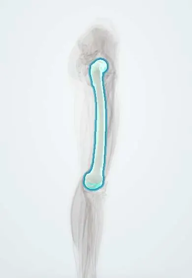
Representative model, for educational purposes only.