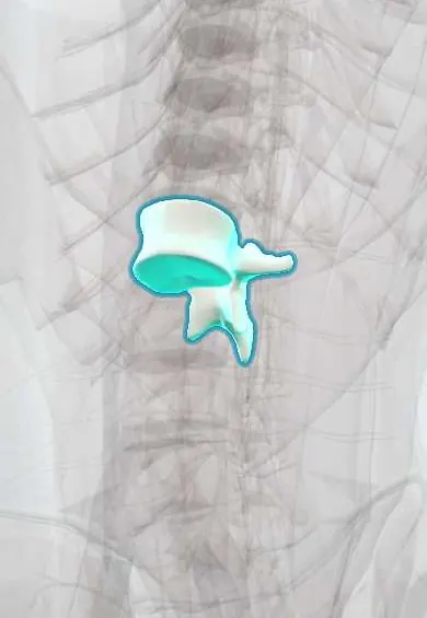
L1 vertebra !
L1 is the first lumbar (lower-back) vertebra, with a large weight-bearing body; the spinal cord ends at about this level.
Official name: L1
Opens in the Durgical app, no sign-up needed.
What it is
The L1 lower-back vertebra, with a large body built to carry weight. The spinal cord ends at about this level.
What it does
Carries the weight of the upper body and, with the disc and facet joints, lets the lower back bend forward and back.
Key facts
- Joints with
- T12 above and L2 below (intervertebral discs, facet joints)
- Landmarks
- Large kidney-shaped body; no costal facets or transverse foramina; mammillary processes; transpyloric plane; conus medullaris ends near L1–L2
Relations
- In front
- Crura of the diaphragm on each side of the body; the abdominal aorta with the superior mesenteric artery leaving at about L1; the cisterna chyli to the right of the aorta; the inferior vena cava farther right
- Lateral
- Psoas major starts from the bodies and discs; the kidneys lie in front of the transverse processes, their hila near this level
- Behind
- Broad, horizontal spine under the thoracolumbar fascia; erector spinae and multifidus in the groove beside it
- Contents
- Conus medullaris with the sacral cord segments, and the start of the cauda equina
How to find it
The spine is counted up from L4 on the iliac crest line, or down from the twelfth rib. In front, the transpyloric plane sits midway from the jugular notch to the top of the pubic symphysis.
On imaging
MRI shows where the conus ends; a conus below L2 in a child suggests a tethered cord. On CT, burst fractures here are judged by canal narrowing and loss of body height.
Common problems
- Disc degeneration and herniation at L4-L5, the most common level for back and leg pain.
- Degenerative spondylolisthesis: L4 slips forward on L5, often with stenosis (more common in women over 50).
- Spinal stenosis: the canal narrows from disc bulging, facet arthritis and a thick ligamentum flavum.
- Compression fractures from osteoporosis or injury.
How it is diagnosed
X-rays (standing, and bending forward and back) show alignment and slipping. MRI shows discs, nerves and stenosis. CT shows bone detail.
Treatment
- Most back pain improves with staying active, physical therapy and pain medicine.
- Stenosis with leg pain when walking that does not improve: decompression (laminectomy).
- Slipping with instability: decompression plus fusion with screws, rods, a cage and bone graft.
Clinical notes
- Conus syndrome versus cauda equina syndrome: the conus gives early, symmetric bladder and bowel loss and saddle numbness with mixed upper and lower motor neuron signs; the cauda equina gives asymmetric root pain and lower motor neuron weakness
- Tethered cord: the conus sits abnormally low, held by a thick filum terminale or a lipoma; pain, gait changes and bladder problems appear with growth
Development
At birth the cord ends lower, at about L2–L3, and reaches its adult level in early infancy because the spine grows faster than the cord.
Related conditions
- Multiple myeloma
- Osteoporotic vertebral compression fracture
- Spinal tuberculosis (Pott disease)
- Tuberculosis
- Osteoporosis
- Malignant spinal cord compression
Test yourself
Think of the answer, then tap the question to see it.
At which level does the adult spinal cord end, and what does an L1 burst fracture risk?
At about the L1–L2 disc; an L1 burst fracture can damage the conus medullaris (conus syndrome) rather than single roots.
Which structures lie on the transpyloric plane at the L1 vertebra?
The pylorus, the neck of the pancreas, the renal hila, the origin of the superior mesenteric artery, the gallbladder fundus and the ninth costal cartilages.
How does conus syndrome at the L1 vertebra differ from cauda equina syndrome?
Conus: early, symmetric bladder and bowel loss and saddle numbness with mixed upper and lower motor neuron signs. Cauda equina: asymmetric root pain and lower motor neuron weakness.
What does a conus medullaris ending below L2 in a child suggest?
A tethered cord, held low by a thick filum terminale or a lipoma.
Educational information, not medical advice. For any symptom, see a health professional.
Its names
- Official Latin (TA2)
- vertebra lumbalis I TA2 1068
- Official Spanish (DeCS)
- Vértebra lumbar L1 DeCS D008159
- English (TA2)
- L1
Related
- T12–L1 disc
- L1–L2 disc
- Lumbar arteries
- Anterior longitudinal ligament
- Endplates
- Low-back facet joints
- Mid-back facet joints
- Interspinales
Sources: Terminologia Anatomica (TA2); DeCS; Anatomy, Back, Lumbar Spine.
Spotted a mistake?We are always improving for the community. Tell us what is wrong and our team will review it.Report a mistake
Representative model, for educational purposes only.