Conditions · Vitamins and minerals
Rickets and osteomalacia !
Bone fails to mineralise for lack of vitamin D, calcium or phosphate: in children it bends (rickets), in adults it softens and hurts (osteomalacia).
Updated 2026-11-01
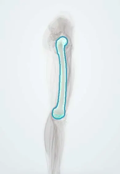
Structures involved
Tap one to see what it is and open it in 3D.
In biochemistry
- Alkaline phosphatase (ALP) as a lab test
- Nuclear receptors
- Vitamin D (calcitriol)
- Calcium and phosphate
In histology
In physiology
- Calcium regulation: PTH, vitamin D and calcitonin
- Bone remodeling
- Phosphate and FGF-23
- Absorption of iron, calcium and vitamin B12
- Vitamins and minerals: a physiological overview
Why it happens
What it is
Defective mineralisation of bone matrix (osteoid). In children, with open growth plates, it is called rickets; in adults, osteomalacia.
Who gets it
It has returned in exclusively breastfed infants without supplements, with dark skin or little sun, and in institutionalised older adults. In tropical areas such as Colombia it is less common thanks to sunlight, but it is seen with malnutrition, malabsorption and kidney disease.
Causes
- Vitamin D deficiency: little sunlight, poor diet, malabsorption, liver disease
- Chronic kidney disease (no 1α-hydroxylation)
- Very low calcium diet
- Renal phosphate wasting: X-linked hypophosphataemia (high FGF23), Fanconi syndrome
- Vitamin D-dependent rickets type 1 (CYP27B1) and type 2 (VDR receptor)
- Drugs: anticonvulsants, antiretrovirals such as tenofovir
Risk factors
- Prolonged exclusive breastfeeding without vitamin D supplements
- Dark skin, full-body clothing, staying indoors
- Prematurity
- Bariatric surgery, coeliac disease
Pathophysiology
Without calcitriol little calcium is absorbed; PTH rises to hold calcium and wastes phosphate in urine. The calcium × phosphate product stays low and osteoid does not mineralise. In the growth plate hypertrophic chondrocytes pile up uncalcified: metaphyses widen and cup and bones bend under weight. In adults soft osteoid builds up: bone pain, muscle weakness and pseudofractures.
Classification
- Calcipenic: insufficient vitamin D or calcium (high PTH)
- Phosphopenic: renal phosphate wasting (normal PTH, high FGF23 in the X-linked form)
- Vitamin D-dependent rickets types 1 and 2
Symptoms and signs
- Children: bowed legs (genu varum) or knock knees, late walking, late fontanelle closure
- Rachitic rosary (swollen costochondral junctions), widened wrists and ankles
- Craniotabes, frontal bossing, late tooth eruption and caries
- Adults: diffuse bone pain, proximal muscle weakness, waddling gait
- Fractures with minor trauma
Diagnosis
- Clinical
- Typical deformities and risk factors in a child; bone pain, proximal weakness and fragility fractures in an adult.
- Tests
- High alkaline phosphatase, low phosphate, low or normal calcium, high PTH, low 25-OH-D in nutritional forms; urine phosphate and FGF23 in phosphopenic forms.
- Imaging
- Wrist or knee X-ray: widened, cupped, frayed metaphyses. In adults, Looser zones (pseudofractures) in the femoral neck, pubis and ribs; low bone density.
Treatment
- Medical
- Replace vitamin D and ensure enough dietary or supplemental calcium
- Medical
- In renal or genetic causes: calcitriol or active analogues, phosphate or specific treatment
- Follow-up
- Follow calcium, phosphate, alkaline phosphatase, PTH and X-rays until healing
- Surgical
- Corrective osteotomy for severe persistent deformities
- Lifestyle
- Sensible sun exposure and vitamin D-containing foods
Complications
- Permanent bone deformity and short stature
- Narrow pelvis that complicates childbirth
- Fractures
- Hypocalcaemic seizures
Prognosis
The nutritional form heals within weeks to months with treatment and many mild deformities correct with growth; genetic forms need lifelong treatment.
Prevention
- Vitamin D supplements for breastfed infants as guidelines advise
- Reasonable sun exposure, fortified foods
- Monitoring people with malabsorption or on anticonvulsants
Test yourself
Think of the answer, then tap the question to see it.
Why does vitamin D deficiency lower serum phosphate?
Low calcium raises PTH, which increases renal phosphate excretion.
What is the rachitic rosary?
Palpable swellings at the costochondral junctions from accumulated unmineralised cartilage and osteoid.
What are Looser zones?
Pseudofractures: radiolucent bands perpendicular to the cortex, typical of osteomalacia.
What separates phosphopenic from calcipenic rickets?
In phosphopenic rickets PTH and calcium are normal and the kidney wastes phosphate (for example, high FGF23 in the X-linked form).
What is the most sensitive biochemical marker of active rickets?
Raised alkaline phosphatase, reflecting increased osteoblast activity.
Related conditions
Educational information, not medical advice. For any symptom, see a health professional.
Sources: Rickets: MedlinePlus Medical Encyclopedia (MedlinePlus, US National Library of Medicine, 2026); Osteomalacia: MedlinePlus Medical Encyclopedia (MedlinePlus, US National Library of Medicine, 2026); Vitamin D Deficiency: MedlinePlus (MedlinePlus, US National Library of Medicine, 2026); Vitamin D-dependent rickets: MedlinePlus Genetics (MedlinePlus, US National Library of Medicine, 2026); Vitamin D Test: MedlinePlus Medical Test (MedlinePlus, US National Library of Medicine, 2026); Textbook of Biochemistry for Medical Students, 6th ed. (Jaypee Brothers Medical Publishers (Vasudevan DM, Sreekumari S, Vaidyanathan K), 2011).
Spotted a mistake?We are always improving for the community. Tell us what is wrong and our team will review it.Report a mistake
Representative model, for educational purposes only.