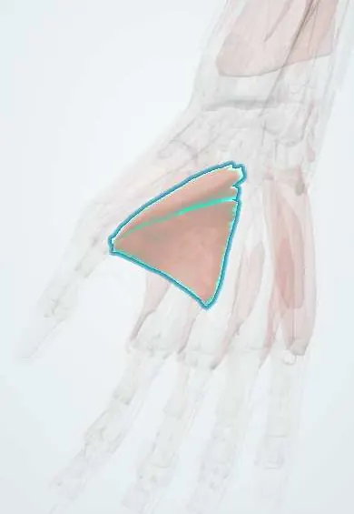
Adductor pollicis !
The adductor pollicis is a fan-shaped muscle deep in the palm near the thumb web; it pulls the thumb toward the palm for a strong pinch or grip.
Opens in the Durgical app, no sign-up needed.
What it is
A fan-shaped muscle deep in the web between thumb and palm.
What it does
Pulls the thumb toward the palm for a strong pinch or grip.
Key facts
- Origin
- Oblique head: capitate and bases of the 2nd–3rd metacarpals; transverse head: shaft of the 3rd metacarpal
- Insertion
- Medial base of the proximal phalanx of the thumb
- Action
- Adducts the thumb (key pinch)
- Nerve
- Deep branch of the ulnar nerve (C8–T1)
- Blood supply
- Deep palmar arch
Relations
- Superficial
- The long flexor tendons to the index and middle fingers and the first two lumbricals; the FPL tendon runs along its lateral edge
- Behind
- First dorsal interosseous, which forms the thumb web with it
- Contents
- Between its oblique and transverse heads, the radial artery as it becomes the deep palmar arch; the deep branch of the ulnar nerve runs with the arch and ends in it
- Lateral
- At its insertion, the ulnar sesamoid of the thumb and the adductor aponeurosis over the thumb UCL
How to find it
Gripping a sheet of paper between the thumb and the side of the index makes it bulge on the palmar side of the first web space; the first dorsal interosseous bulges on the back.
Examination
- Froment sign: flexion of the thumb IP joint while holding a sheet of paper in a key pinch indicates weak adductor pollicis (ulnar nerve palsy)
- Jeanne sign: hyperextension of the thumb MCP joint during the same pinch, the same meaning
On imaging
Its aponeurosis is the key landmark in skier's thumb. When the ulnar collateral ligament of the thumb MCP joint tears, the torn end can flip above the aponeurosis (Stener lesion) and will not heal without surgery. Ultrasound or MRI shows the balled-up stump above the aponeurosis edge. Radiographs first look for an avulsion fragment of the proximal phalanx base.
Clinical notes
- Stener lesion after a skier's thumb injury: the adductor aponeurosis lies between the torn ligament and its insertion, usually an indication for surgery
- Weak key pinch is one of the most disabling losses after ulnar nerve injury and a common target for tendon transfer
Anatomical variants
- Some texts count it with the thenar muscles, others apart because of its ulnar supply; each of its two heads is built of several fascicles
Development
Its motor supply comes from the C8–T1 segments, so a lower brachial plexus birth injury (Klumpke palsy) weakens it along with the other intrinsic hand muscles, leaving a clawed hand and a weak pinch.
Related conditions
Test yourself
Think of the answer, then tap the question to see it.
What is the Froment sign?
In ulnar nerve palsy, the thumb tip bends when gripping a sheet of paper against the index, because FPL replaces the weak adductor pollicis.
What is a Stener lesion, and how is the adductor pollicis involved?
The torn thumb MCP ulnar collateral ligament flips above the adductor aponeurosis, which then lies between ligament and insertion, so it cannot heal on its own.
What nerve supplies the adductor pollicis, and which vessel passes between its heads?
The deep branch of the ulnar nerve; the radial artery enters the palm between its heads to form the deep palmar arch.
Educational information, not medical advice. For any symptom, see a health professional.
Its names
- Official Latin (TA2)
- adductor pollicis TA2 2526
- Official Spanish (DeCS)
- Músculo aductor del pulgar
- English (TA2)
- Adductor pollicis
Related
- Thumb UCL
- Ulnar nerve in the hand
- Deep palmar arch
- Capitate
- 1st metacarpal
- 2nd metacarpal
- 3rd metacarpal
- Proximal phalanx, thumb
Sources: Terminologia Anatomica (TA2); Anatomy, Shoulder and Upper Limb, Hand Adductor Pollicis.
Spotted a mistake?We are always improving for the community. Tell us what is wrong and our team will review it.Report a mistake
Representative model, for educational purposes only.