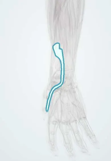
FPL tendon !
The flexor pollicis longus tendon runs through the carpal tunnel to the thumb tip; it is the only tendon that bends the thumb's end joint.
Official name: Flexor pollicis longus tendon
Opens in the Durgical app, no sign-up needed.
What it is
The thumb's long flexor tendon, the most thumb-side tendon in the carpal tunnel.
What it does
Bends the tip of the thumb.
Key facts
- Attaches
- Most radial tendon in the carpal tunnel, in its own sheath (radial bursa), between the two heads of flexor pollicis brevis to the base of the distal phalanx of the thumb
- Function
- The only flexor of the thumb's interphalangeal joint; with the FDP of the index it makes the 'OK' sign that tests the anterior interosseous nerve
- Tight in
- Thumb extension with wrist extension
Relations
- Course
- From the front of the radius its tendon runs through the carpal tunnel, between the two heads of the flexor pollicis brevis, then under the thumb's A1, oblique and A2 pulleys to the distal phalanx
- Superficial
- Flexor retinaculum, with the median nerve just in front and to its ulnar side in the carpal tunnel
- Deep
- Pronator quadratus and the distal radius at the watershed line, then the scaphoid and trapezium in the floor of the carpal tunnel
- Lateral
- FCR tendon, in its own tunnel through the trapezium, outside the carpal tunnel
- Medial
- Index finger FDS and FDP tendons in the carpal tunnel
How to find it
With the proximal phalanx of the thumb held still, bending the tip of the thumb shows the tendon working; it can be felt crossing the palmar crease of the thumb's MCP joint.
Examination
- Isolated FPL test: the thumb's proximal phalanx is held and IP flexion is asked for; none means the tendon is cut or the anterior interosseous nerve is not working
- 'OK' sign: a flat pinch with straight thumb IP and index DIP points to an anterior interosseous nerve palsy; if only the thumb is affected, a ruptured FPL is more likely
On imaging
Ultrasound shows continuity, fraying over a plate edge and the retracted stump. On the lateral X-ray, a volar plate standing proud beyond the watershed line of the distal radius is a warning sign.
Clinical notes
- Volar plating of distal radius fractures: a plate placed too far distally or standing proud can rub through the tendon weeks to months later
- Rupture in rheumatoid arthritis over a scaphoid osteophyte (Mannerfelt–Norman lesion)
- Horseshoe abscess: infection spreading between the radial and ulnar bursae through the space of Parona
Anatomical variants
- An accessory head (Gantzer's muscle), from the medial epicondyle or coronoid process, may compress the anterior interosseous nerve
- A tendinous link between FPL and the index FDP (Linburg–Comstock) makes the index tip bend when the thumb flexes
Test yourself
Think of the answer, then tap the question to see it.
What does the FPL tendon flex, and what is its position in the carpal tunnel?
It is the only flexor of the thumb IP joint and the most radial tendon in the carpal tunnel, in its own sheath (radial bursa).
What is a horseshoe abscess involving the FPL tendon sheath?
Infection spreading between the radial bursa (FPL sheath) and the ulnar bursa through the space of Parona.
Which fracture implant most often frays the FPL tendon?
A volar plate for a distal radius fracture placed beyond the watershed line or standing proud.
What is the Mannerfelt lesion?
Rheumatoid rupture of the FPL tendon over a scaphoid spur (osteophyte).
Educational information, not medical advice. For any symptom, see a health professional.
Its names
- Official Latin (TA2)
- tendo musculi flexoris longi pollicis TA2 2492
- Official Spanish (DeCS)
- Tendón del músculo flexor largo del pulgar
- English (TA2)
- Flexor pollicis longus tendon
Related
- Median nerve
- Pronator quadratus
- FPL
- 1st metacarpal
- Distal phalanx, thumb
- Proximal phalanx, thumb
- Radius
- Thumb sesamoids
Sources: Terminologia Anatomica (TA2); Anatomy, Shoulder and Upper Limb, Hand Flexor Pollicis Longus Muscle; Anatomy, Shoulder and Upper Limb, Hand Long Flexor Tendons and Sheaths.
Spotted a mistake?We are always improving for the community. Tell us what is wrong and our team will review it.Report a mistake
Representative model, for educational purposes only.