Endocrine system · Head and neck
Pineal gland !
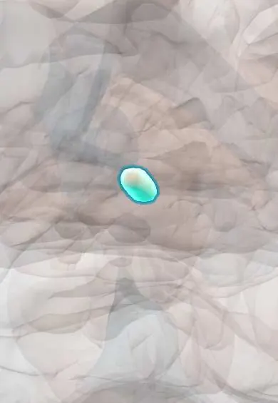
The pineal gland is a tiny pine-cone-shaped gland deep in the brain; it makes melatonin, which helps set the sleep cycle.
Opens in the Durgical app, no sign-up needed.
What it is
A tiny pine-cone gland at the back of the third ventricle.
What it does
Makes melatonin, which helps set the sleep cycle.
Key facts
- Where
- Behind the third ventricle, above the superior colliculi
- Blood supply
- Posterior choroidal arteries
- Nerve
- Sympathetic (light information via the retina and hypothalamus)
- Function
- Melatonin (sleep-wake rhythm)
Relations
- In front
- Back wall of the third ventricle, with the pineal recess; its stalk carries the habenular commissure above and the posterior commissure below
- Below
- Superior colliculi of the midbrain and the opening of the cerebral aqueduct beneath them
- Above
- Splenium of the corpus callosum and the internal cerebral veins, which join above it
- Behind
- Quadrigeminal cistern with the great cerebral vein, which drains into the straight sinus; the cerebellar vermis below and behind
How to find it
Not palpable. On CT its calcification marks the midline just behind the third ventricle, above the superior colliculi; surgeons reach the region from behind, for example between the cerebellum and the tentorium (supracerebellar infratentorial approach).
Examination
- Upward gaze and pupil checks: asking the person to look up and comparing pupil reactions to light and to a near target; upgaze palsy with light–near dissociation suggests a pineal region mass pressing on the tectum
On imaging
CT shows calcification in most adults; calcification in a young child is unusual and raises concern for a tumour. Sagittal MRI is the view for pineal masses and for small fluid-filled pineal cysts, a frequent incidental finding that is usually harmless.
Clinical notes
- Germinoma is the most common pineal tumour and occurs mainly in boys and young men; blood and CSF tumour markers help separate germ cell tumours from other types
- Hydrocephalus from a pineal mass is relieved by a ventricular shunt or an endoscopic opening of the third ventricle floor
- On CT or older skull radiographs, a calcified pineal pushed away from the midline points to a mass or swelling in one hemisphere
- Germ cell tumours of the pineal region that secrete hCG can cause precocious puberty in boys
Anatomical variants
- Small pineal cysts are common incidental findings on MRI and are usually harmless
Development
It grows as an outpouching of the roof of the diencephalon (epithalamus), at the back of the third ventricle.
Test yourself
Think of the answer, then tap the question to see it.
What hormone does the pineal gland make, and how does light affect it?
Melatonin; darkness raises it and light suppresses it, via the retina, suprachiasmatic nucleus and sympathetic fibres from the superior cervical ganglion.
What is Parinaud syndrome and what causes it?
Loss of upward gaze, pupils reacting to near but not light, lid retraction and convergence-retraction nystagmus, from a pineal region tumour pressing on the tectum.
Why can a pineal tumour cause obstructive hydrocephalus?
It squeezes the cerebral aqueduct beneath the pineal gland.
Which is the most common pineal tumour, and in whom?
Germinoma, mainly in boys and young men.
Educational information, not medical advice. For any symptom, see a health professional.
Its names
- Official Latin (TA2)
- glandula pinealis TA2 3862
- Official Spanish (DeCS)
- Glándula Pineal DeCS D010870
- English (TA2)
- Pineal gland
- Also called
- epiphysis cerebri · pineal body
Sources: Terminologia Anatomica (TA2); DeCS; Physiology, Pineal Gland; Tratado de anatomía humana (9.ª ed.), tomo II, Libro V, cap. VI: Cerebro (configuración exterior e interior, ventrículos, formaciones coroideas, epífisis).
Spotted a mistake?We are always improving for the community. Tell us what is wrong and our team will review it.Report a mistake
Representative model, for educational purposes only.