Cardiovascular system · Head and neck
Anterior communicating artery !
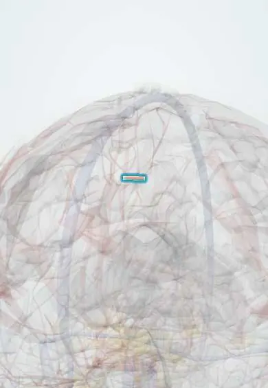
The anterior communicating artery is a short link joining the two anterior cerebral arteries; it is the most common site of brain aneurysms.
Opens in the Durgical app, no sign-up needed.
What it is
A very short bridge joining the two anterior cerebral arteries.
What it does
Closes the front of the circle of Willis; the most common site of brain aneurysms.
Key facts
- Origin
- Joins the two anterior cerebral arteries
- Branches
- Small perforators
- Supplies
- Closes the front of the circle of Willis; most common aneurysm site
Relations
- Course
- A short bridge across the midline in front of the optic chiasm, joining the two anterior cerebral arteries where their A1 and A2 segments meet
- Below
- Optic chiasm
- Behind
- Lamina terminalis and the front of the hypothalamus
- Above
- Gyrus rectus and the medial orbital surface of the frontal lobes
How to find it
It cannot be felt. It lies just above the optic chiasm, in front of the lamina terminalis, joining the two A1 segments; in a pterional craniotomy it is reached by following the internal carotid and A1 across the chiasm, often after removing a little of the gyrus rectus.
On imaging
CT angiography finds most aneurysms here; catheter angiography, with angled views that open up the junction, plans treatment.
Common problems
- Brain aneurysm: a weak bulge in the artery wall, most often at this junction.
- Rupture causes a subarachnoid haemorrhage: a sudden 'worst headache of my life'.
How it is diagnosed
CT and CT angiography; angiography with a catheter to plan treatment; unruptured aneurysms are often found by chance on scans.
Treatment
- Clipping: a small metal clip across the neck of the aneurysm through a craniotomy.
- Coiling: soft platinum coils packed inside it through a catheter from the groin or wrist.
- Watching some small unruptured aneurysms, with blood pressure control and no smoking.
Clinical notes
- Ruptured aneurysms are secured by endovascular coiling or surgical clipping, chosen by the shape and position of the aneurysm
- A large aneurysm can press on the optic chiasm or optic nerve and cause visual field loss
- Days after the bleed, vasospasm can narrow both anterior cerebral arteries
Anatomical variants
- It may be duplicated, fenestrated or missing, with the two anterior cerebral arteries fused at that point
Related conditions
Test yourself
Think of the answer, then tap the question to see it.
What is the most common site of berry aneurysms in the circle of Willis?
The anterior communicating artery, which joins the two anterior cerebral arteries.
Where does a ruptured anterior communicating artery aneurysm bleed on CT?
Into the interhemispheric fissure and the gyrus rectus.
What can damage to the perforators of the anterior communicating artery cause?
Memory loss, confabulation and personality change, from injury to the basal forebrain.
How are ruptured anterior communicating artery aneurysms secured?
By endovascular coiling or surgical clipping, chosen by the shape and position of the aneurysm.
Educational information, not medical advice. For any symptom, see a health professional.
Its names
- Official Latin (TA2)
- arteria communicans anterior TA2 4517
- Official Spanish (DeCS)
- Arteria comunicante anterior
- English (TA2)
- Anterior communicating artery
Related
Sources: Terminologia Anatomica (TA2); Neuroanatomy, Circle of Willis.
Spotted a mistake?We are always improving for the community. Tell us what is wrong and our team will review it.Report a mistake
Representative model, for educational purposes only.